4.7 Image Processing by Belkousov-Zhabotinsky-Type Media
|
4.7 Image Processing by Belkousov-Zhabotinsky-Type Media
Wave algorithms in image processing have been discussed in literature beginning from the late 1960s (see, for instance, Pratt 1978). They were "prairie fires" and some other versions that were promising for efficient determination of shape description due to the high parallelism inherent in them.
The next step in the further development of wave-image processing approaches was based on the unique properties of reaction-diffusion systems. Both high parallelism and nonlinear dynamic mechanisms that increased behavioral complexity were the basis behind the elaboration of image processing methods.
The foundations of information processing by chemical light-sensitive reactiondiffusion media were laid in pioneering studies performed by Lothar Kuhnert (1986, 1986b), which were followed by the paper "Information processing using lightsensitive waves", published by Kuhnert, Agladze, and Krinsky (1989). This was an important study that determined the physical and chemical principles inherent in information processing by reaction-diffusion media. At the same time, the information processing basis of this work remained meager. Detailed investigations of information processing capabilities in chemical light-sensitive reaction-diffusion media were performed in the beginning of the 1990s (see Rambidi and Maximychev 1997 and references therein). It became clear as a result of these investigations that fundamental differences exist between:
-
Image processing of positive and negative images of initial pictures
-
responses of the medium to the light excitations and information processing operations performed by the medium based on these responses
-
information processing of black-and-white pictures, pictures having several levels of brightness, and halftone pictures
Let us define the positive image of a picture as the image corresponding to a typical picture inherent to human surroundings. If the notion of "typical picture" is uncertain (e.g., the case of geometrical figures; see, for instance, figure 4.10), let us define the positive image as a dark figure on a light background. Black-and-white and halftone pictures will be used below. Images of these pictures could be considered as a set of optical density values D(i) corresponding to each point of the picture (0 < D(i) < D(∞), where D(∞) is the maximum value of the optical density). The negative image of the picture is defined as a set of inverted density values (DNi = D∞ Di). Adobe Photoshop software was used for the positive-negative transformations.
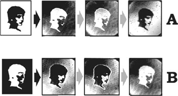
Figure 4.10: Processing positive (A) and negative (B) black and white pictures by a Belousov-Zhabotinsky medium. Here and in following figures black arrows correspond to input of the initial picture, grey ones show transformation of the image by the medium. The sequences of images (A and B) in this figure and in figures 4.3 and 4.5 correspond to one period of Belousov-Zhabotinsky medium oscillation. This period was about 20–30 sec. Time intervals between separate images throughout of each period were 3–5 sec.
There were two reasons why this work was performed recently. The first of these was the advent of new experimental techniques during the last decade that have proved to be indispensable for the investigations of complex reaction-diffusion dynamics. The designing of reaction-diffusion media optimal for solving a problem under investigation forms the basis of this technique. The most remarkable features of this technique lie in employing modern polymeric materials as specialized matrices for the medium and immobilization of separate chemical components of the reaction-diffusion system. As a result, improved techniques gave us the opportunity to sufficiently increase the reliability of data in comparison with the data of Kuhnert, Agladze, and Krinsky (1989) and that of subsequent investigations (Rambidi and Maximychev 1997).
Understanding that the experimental data formerly obtained was incomplete and even somewhat incompatible was the second reason why this work was performed recently. New important regimes of image processing by reaction-diffusion media have been found as a result of investigations based on the improved technique, enabling one to explain qualitatively the whole set of the experimental data using the simple FKN model (Field and Burger 1985).
There are four basic points that determine the character of the image processing by light-sensitive reaction-diffusion media (see details in Rambidi and Maximychev 1997). They are:
-
The composition of the Belousov-Zhabotinsky reagent
-
Positive or negative versions of the initial picture
-
The optical contrast of the picture
-
Exposure in the process of initial picture input into the medium
The Case of a Black-and-White Picture
The negative image of the initial picture is revealed after its input into the medium. After that, the following stages of image evolution can generally be observed:
-
Contour enhancement
-
Spatial evolution of the contour (the shrinkage of the contour or its spreading, see Rambidi and Maximychev 1997)
-
Appearance of the positive image of the input picture
(Some of the above steps are optional, depending on operational conditions.)
Basic stages of picture evolution for positive and negative images of the input picture are shown in figure 4.10.
This process can be qualitatively explained based on the simplest FKN model for stirred Belousov-Zabotinsky-type medium (see, for instance, Field and Burger 1985).
According to this model, the medium can be described by two ordinary differential equations (ODE) corresponding to activator and inhibitor dynamics. Stationary states of these equations could be characterized by a set of null clines. Light-sensitive Belousov-Zhabotinsky media functioning in oscillating mode were used for the investigation of the discussed image processing capabilities. The null clines corresponding to this mode are shown in figure 4.11. One of them (S-type) corresponds to inhibitor ODE, while the other corresponds to activator ODE. The main feature of this oscillating mode is that null clines have no common stable point.
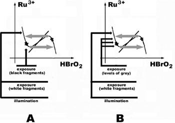
Figure 4.11: Schematic representation of image processing mechanism inherent in a light-sensitive BelousovZhabotinsky medium functioning in oscillating mode. Null clines and mechanisms of image transformation are given for cases of black and white (A) and half-tone (B) pictures. See also explanations in the text.
In a Belousov-Zhabotinsky-type reaction proceeding in oscillating mode, the color of the reagent changes between orange (high concentration of Ru2+ and low concentration of Ru3+) and fluorescent bluish (high concentration of Ru3+ and low concentration of Ru2+). A black-and-white video camera was used to record the medium evolution (using a blue filter to increase the contrast of the image). The color of the records input into computer memory thus varied from black to white (high and low concentrations of Ru2+, see figure 4.11).
The typical procedure of the experiment was as follows:
First, the medium was illuminated by white light to remove traces of previous experiments. The illumination initiated the photochemical reactions, which sharply increased the concentration of Ru3+ (light color). At the same time, the inhibitor (HBrO2) concentration was expected to decrease according to the FKN model. After that, a picture under consideration was projected onto the surface of the medium. As a result:
-
The concentration of Ru3+ remained high in the areas of the medium where white fragments of the initial picture were projected (the upper part of the left null cline branch).
-
The concentration of Ru3+ decreased and concentration of Ru2+ increased in the areas of the medium where black fragments of the picture were projected (the lower part of the left null cline branch).
A faint positive image of the input picture appeared, usually just after switching off the exposure. At that point, an oscillating Belousov-Zhabotinsky-type process began in the medium. The character of the medium evolution could be described as moving points corresponding to concentrations in exposed areas along null clines toward unstable foci (see figure 4.11). The basic feature of this movement was the tendency to decrease concentration of Ru3+ in white exposed fragments and to increase concentration of Ru3+ in black exposed fragments of the medium (here the point jumps to the right branch of the null cline and moves upward). Therefore the image of the input picture turned into a negative one. In the process of the following evolution, a negative image would turn into a positive one, and so on.
Basic responses of a Belousov-Zhabotinsky medium to light excitation and image processing operations based on these responses have already been discussed in detail earlier (Rambidi and Maximychev 1997). Image processing operations performed by active chemical media have proved to be similar to human visual capabilities. There are two main sets.
The first can be defined as "description of the general features of an object". This set includes such primitive operations as concentration on the general outline of an image, removing small immaterial features, "addition to the whole" operations, and, in particular, restoration of an image with defects.
The second set of image processing operations can be determined as "switching to the details of an image". It includes contour enhancement, segmentation (i.e., division of an image into simple parts), image skeletonizing, and italicizing small features of an image.
In this case, the main factors controlling the character of the image evolution are the negative or positive form of the initial picture, and exposure of the light excitation. These two factors are important to control the contour-enhancement mode. Decreasing pH and increasing exposure determine conditions for revealing this mode (see details and examples in Rambidi and Maximychev 1997).
We mention here that basic features of image processing by Belousov-Zhabotinsky media were first discussed in Kuhnert, Agladze, and Krinsky (1989).
The Case of a Halftone Initial Picture
The transformation of positive and negative pictures is shown in figure 4.12. The halftone picture is transformed first by the reaction-diffusion medium into a blackand-white form.
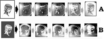
Figure 4.12: Processing positive (A) and negative (B) half-tone pictures by a Belousov-Zhabotinsky medium. See also caption to figure 4.10.
The positive input picture is transformed into its negative black-and-white image step by step—beginning from its darkest fragments. The contour enhancement mode then appears and in the end of the transformation period, a halftone positive image is observed.
In the case of a negative input picture, the main part of the brightest fragments is revealed simultaneously as a black-and-white positive image at the very beginning of the image evolution. After that, more and more dark fragments are enhanced step by step in positive black-and-white forms of the input picture.
This complicated evolution of a halftone image has not been observed before. Although the simple FKN model does not explain all details of the image evolution, it is possible to conclude only that fragments of different brightness should appear simultaneously step by step in the process of image evolution.
When the picture is projected on the layer of Ru-based Belousov-Zhabotinsky reagent, the information on brightness of the picture is introduced into the medium. It is stored as a spatial distribution of the reaction component's concentrations. In this case, the section of the null cline corresponding to different levels of gray spreads along its left branch (figure 4.11.). In the process of the image evolution in the medium (moving along the null cline), the darkest fragments of the stored image emerge first and then step by step all others with less concentration of Ru+3. This process virtually visualizes information about the brightness of the initial picture, transforming it from a spatial into a temporal form.
All control factors that determine the processing of black-and-white pictures work in the case of halftone images. But one more factor is important in this case—the optical contrast within the input picture. For instance, changing the contrast of the initial negative picture gives the opportunity to enhance the contour of the whole picture (figure 4.13, c1) and its separate parts (figure 4.13, c2)
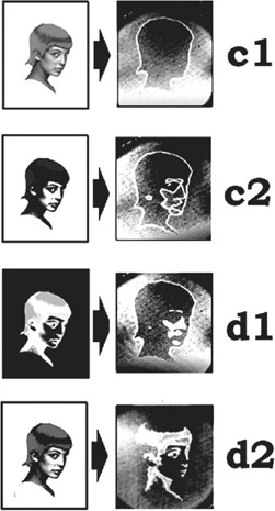
Figure 4.13: Different variants of half-tone picture processing: c1, contour enhancement (low contrast of the initial picture); c2, contour enhancement (high contrast of the initial picture); d1, enhancement of the basic shape of the initial picture; d2, enhancement of details of the initial picture.
Information processing modes of chemical reaction-diffusion media open new ways that seem to be important for practical applications.
There is a great variety of responses of Belousov-Zhabotinsky media to the photochemical input of pictures having several levels of brightness and halftone pictures. These responses allow one to solve many image processing and image recognition problems. Let us consider several examples:
The processing of a negative initial picture leads to enhancement of the basic picture shape (figure 4.13, d1), while the processing of a positive picture gives the possibility to accentuate fine features of the picture (figure 4.13, d2).
The consecutive enhancement of fragments with different levels of brightness is an important feature of the image processing. The evolution of a picture with five levels of brightness in a reaction-diffusion medium is shown in figure 4.14. Numbers in the initial picture of this figure correspond to relative optical densities (0 for white and 100 for black). The separate enhancement of all fragments of the picture indicates an effective enough transformation function of the medium (level of darkness versus consecutive enhancement).
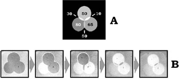
Figure 4.14: Enhancement of picture fragments having different brightness by a Belousov-Zhabotinsky medium. A and B are the initial picture and subsequent results. Numbers in the initial picture correspond to relative optical densities of the image fragments. See also caption to figure 4.10.
The interpretation of aerial and satellite pictures is one of the most widespread and important problems among different image processing tasks. In practical terms, it comes down to dividing the picture into fragments with specific levels of brightness. It was shown earlier that reaction-diffusion media prove to be promising instruments for solving this problem (Rambidi and Maximychev 1997). The new experimental technique elaborated broadens these possibilities of reaction-diffusion media.
The consecutive enhancement of fragments with different levels of brightness enables one to restore individual components of the picture where these components overlap. This process is shown in figure 4.15. The whole overlapped figure (p1), then the fragment having lower darkness (p2), and finally the overlapped part of the picture (p3) appear consecutively in the course of the image evolution. Using standard image processing operations performed by a conventional digital computer gives the opportunity to restore the second component of overlapped picture based on these experimental data (f1, f2).
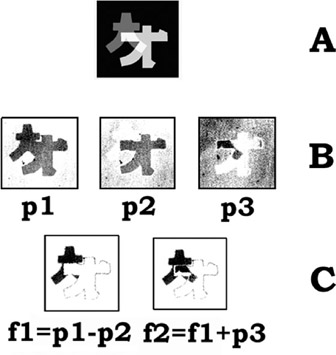
Figure 4.15: The restoration of individual components of the picture where components overlap. Initial picture (A), evolution of the overlapped picture in the reaction-diffusion medium (B), and image processing operations that restore an overlapped component (C).
Figure 4.16 shows the situation of "how many original spots are on the skin of a cat". There are two sets of spots: original spots of the cat (p), and additional spots introduced from outside (p1). In the course of processing the negative image of the picture (n1), the following stages are observed: the whole overlapped picture, then the whole assembly of the spots, and finally the spots from the outside only. It enables one to extract an original image from "rubbish" pictures.
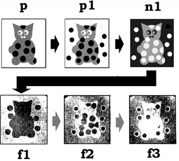
Figure 4.16: How many spots are on the skin of a cat. Original initial picture (p), initial picture together with additional spots statistically distributed (p1), and the negative image of the total picture (n1) are at the top of the figure. Below are: the whole overlapped picture (f1), the whole assembly of spots (f2), and spots introduced from outside (f3).
It should be noted that the processes of the temporal image evolution can be rather successfully described by solutions to the reaction-diffusion equations. For instance, Balkarey, Evtichov, and Elinson (1991) calculated the process of contour enhancement and some other features. The amplifying of small features and spoiled image restoration were demonstrated by Price, Wambacq, and Oosterlinck (1990).
All results discussed above were obtained using light-sensitive BelousovZhabotinsky-type media. (In Jinguji et al. 1995, the formation and propagation of rectangular chemical waves are studied.)
Other chemical reaction systems were used by Adamatzky and Tolmachev (Tolmachev and Adamatzky 1996; Adamatzky and Tolmachev 1997) to perform information processing operations. The reaction between palladium chloride and potassium iodide in a thin layer of agar gel made it possible to compute Voronoi diagrams (Tolmachev and Adamatzky 1996). The formation of Prussian blue in agar gel was used for computation of planar shape skeletons (Adamatzky and Tolmachev 1997).
|
- The Effects of an Enterprise Resource Planning System (ERP) Implementation on Job Characteristics – A Study using the Hackman and Oldham Job Characteristics Model
- Data Mining for Business Process Reengineering
- Intrinsic and Contextual Data Quality: The Effect of Media and Personal Involvement
- Healthcare Information: From Administrative to Practice Databases
- Development of Interactive Web Sites to Enhance Police/Community Relations