 | Note: Large images and tables on this page may necessitate printing in landscape mode.
Copyright 2007 The McGraw-Hill Companies. All rights reserved.
Clinician's Pocket Reference > Chapter 7. Clinical Microbiology >
General Principles of Clinical Microbiology Some of the most common tests performed on patients are the procurement of tissue or body fluids for direct detection of pathogenic organisms to prove or disprove the presence of infection. The results of these tests are critical in guiding the selection of antibiotics for targeted therapy. The following clinical microbiology principles must be considered. Severity or Degree of Risk: There is a difference between an otherwise healthy patient with a complaint of dysuria consistent with a UTI versus a patient with neutropenia and a high fever. The first needs a simple urinalysis with a routine bacterial culture. The second needs a "pan" culture (as in the prefix "pan-," meaning "all" or "every"), which includes a pair of blood cultures, urinalysis with culture and sensitivity, sputum sample if a productive cough is present), and a chest x-ray to rule out pneumonia. The second patient also needs prompt treatment with empiric broad-spectrum antibiotics because she is at high risk of septicemia and death. Broad Coverage with Empiric Antibiotics: Initiation of antibiotics that broadly cover a newly recognized infection in a timely and appropriate manner often is lifesaving. Selecting the wrong antibiotic, the wrong dose, an improper route, or delaying treatment, however, can increase morbidity and mortality. Timing: Whenever feasible, specimens should be obtained and cultures performed before antibiotics are started. However, antibiotics should never be delayed in the face of a possible life-threatening infection, such as meningitis. After the culture data become available, antibiotic therapy can be narrowed or "de-escalated" to the antibiogram of the recovered organism. Source Control: Collections of pus and infected fluids such as abscesses and empyema must be drained if at all possible. Failure to drain pockets of infection can compromise the outcome. The classic example is necrotizing fasciitis, which is a surgical emergency. Without surgery, mortality approaches 100%. True Infection versus Contamination and Colonization: True infection is almost always accompanied by inflammation, usually marked by the presence of neutrophils in clinical specimens (absent in neutropenia) and clinical signs and symptoms. The presence of a large number of epithelial cells in a sample or the growth of normal skin flora often signifies contamination and colonization secondary to improper collection of specimens, although there are exceptions. Antimicrobial Resistance: Drug resistance is a serious problem in modern medicine. In the past medicine stayed ahead of antimicrobial resistance with the development of new antibiotics to overcome new resistance patterns. Now, as vancomycin-resistant enterococci spread throughout the health care system and new clones of methicillin-resistant Staphylococcus aureus become more prevalent, antibiotic resistance is minimized only through proper antibiotic stewardship. To this end the CDC has launched a 12-step program for preventing antimicrobial resistance in hospitals. | CDC Campaign to Prevent Antimicrobial Resistance in Healthcare Settings: 12 Steps to Prevent Antimicrobial Resistance Among Hospitalized Adults |
|---|
| Prevent Infection | | Step 1. Vaccinate: Give influenza/pneumococcal vaccine to at-risk patients before discharge; get influenza vaccine annually. | | Step 2. Get the Catheters Out: Use catheters only when essential; use the correct catheter; use proper insertion and catheter-care protocols; remove catheters when they are no longer essential. | | Diagnose and Treat Infection Effectively | | Step 3. Target the Pathogen: Obtain cultures; target empiric therapy to likely pathogens and local antibiogram; target definitive therapy to known pathogens and antimicrobial susceptibility test results. | | Step 4. Access the Experts: Consult infectious diseases experts about patients with serious infections. | | Use Antimicrobials Wisely | | Step 5. Practice Antimicrobial Control: Engage in local antimicrobial control efforts. | | Step 6. Use Local Data: Know your antibiogram; know your patient population. | | Step 7. Treat Infection, Not Contamination: Use proper antisepsis for blood and other cultures; culture the blood, not the skin or catheter hub; use proper methods to obtain and process all cultures. | | Step 8. Treat Infection, Not Colonization: Treat pneumonia, not the tracheal aspirate. Treat bacteremia, not the catheter tip or hub. Treat UTI, not the indwelling catheter. | | Step 9. Know When to Say "No" to Vanco: Treat infection, not contaminants or colonization. Fever in a patient with an IV catheter is not a routine indication for vancomycin. | | Step 10. Stop Antimicrobial Treatment: When infection is cured; when cultures are negative and infection is unlikely; when infection is not diagnosed. | | Prevent Transmission | | Step 11. Isolate the Pathogen: Use standard infection control precautions. Contain infectious body fluids (follow airborne, droplet, and contact precautions). When in doubt, consult infection control experts. | | Step 12. Break the Chain of Contagion: Stay home when you are sick. Keep your hands clean. Set an example. |
|
(Adapted from http://www.cdc.gov/drugresistance/healthcare/ha/12steps_HA.htm. Accessed July 3, 2006.) |
|
Microbiology Techniques Acid-Fast Stain (AFB Smear, Kinyoun Stain) Clinical microbiology labs can perform a "modified" acid-fast stain for organisms that are weakly acid-fast staining (eg, Nocardia spp.). Acid-fast bacilli (AFB) stain red to bright pink against the light blue background (Mycobacterium tuberculosis [TB], Mycobacterium scrofulaceum, M. avium-intracellulare, and others). These organisms have a beaded rod appearance under oil immersion and must be cultured on specialized media. Rapid-growing AFB include Mycobacterium abscessus, Mycobacterium chelonae, and Mycobacterium fortuitum and can usually be cultured in fewer than 7 d. Most other AFB (M. tuberculosis, M. avium complex, Mycobacterium kansasii, Mycobacterium marinum) take at least 7 10 d to grow. Mycobacterium gordonae is thought to be nonpathogenic. Blood Culture Peripheral cultures are always preferred over cultures drawn through a catheter. All bloodstream catheters become colonized over time, which causes a high rate of false-positive cultures. To help distinguish contamination from true bacteremia, always draw culture specimens in pairs with at least one and preferably both specimens drawn from peripheral blood. Procedure - 1. Review the technique of venipuncture (see Chapter 13). Clean and disinfect the skin first with alcohol and then with chlorhexidine to reduce skin flora contamination. Do not touch the site after cleaning or before needle entry. If the needle is touched or you need to re-enter the vein, discard the needle and start again.
- 2. Culture technique varies from institution to institution. Use a closed vacuum collection system that incorporates the collection bottle (eg, BacTec). Keep the collection bottle below the site to prevent reflux. An aerobic bottle and an anaerobic bottle constitute a "set" drawn from the same venipuncture site.
- 3. The volume of blood drawn influences results, with a 3% increase in sensitivity per each additional milliliter of blood obtained. Collect 10 mL of blood per culture for adults. Usually two or three sets of blood cultures are drawn at intervals of 30 60 min. However, if antibiotics are being started immediately, it is acceptable to draw the cultures closer together.
- 4. Special tubes and culture technique are required for mycobacteria and fungi. Centrifugation lysis (isolator) systems are used to culture M. avium-intracellulare, M. tuberculosis, and dimorphic fungi such as Histoplasma capsulatum. Also perform an acid-fast smear. Contact your laboratory for specific instructions and special tubes.
Interpretation: Preliminary blood culture results are usually available in 12 48 h and should not be formally reported as negative before 4 d. - A single blood culture that is positive for one of the following organisms usually suggests contamination; on rare occasions these agents are the causative pathogen: Staphylococcus epidermidis, Staphylococcus hominis, Bacillus spp., and Corynebacterium diphtheriae (and other diphtheroids).
- Negative results do not exclude bacteremia, and false-positives can result from contamination. Gram-negative organisms, fungi, and anaerobes are considered pathogenic until proven otherwise.
- The presence of yeast in the blood necessitates treatment with a systemic antifungal agent and repeated blood cultures to document clearance. An ophthalmologic exam to rule out endophthalmitis is mandatory. Yeast usually enters the blood through central line infection, PICC line infection, or a GI source. Removal of the catheter with subsequent blood cultures to document the clearance of fungemia is usually mandatory.
- The most common cause of culture-negative infective endocarditis is previous antibiotic therapy. However, a variety of fastidious and difficult to culture pathogens can cause infective endocarditis. The classic group is the so-called HACEK organisms (slow-growing gram-negative bacteria in the oropharynx): Haemophilus parainfluenzae and aphrophilus, Actinobacillus, Cardiobacterium, Eikenella (called the "fight bite bug" after the hand soft-tissue infection sometimes contracted by a person who strikes another person in the mouth), and Kingella. Other causes of culture-negative infective endocarditis are Bartonella spp., Coxiella burnetii (the agent of Q fever), brucellosis, psittacosis, and the causative organism of Whipple disease, Tropheryma whippleii. If a fastidious organism is suspected, the microbiology lab should be informed and cultures held for 4 wk. Special culture media can also be ordered.
Darkfield Examination Used to identify Treponema pallidum, the organism that causes syphilis. Rectal and oral lesions cannot be examined with this technique because of the presence of nonpathogenic spirochetes. Giemsa Stain Used to identify intracellular organisms such as chlamydiae and Plasmodium (malaria) and other parasites Gonorrhea Smear and Culture Neisseria gonorrhoeae can be cultured from many sites, including female genital tract (endocervix the preferred site), male urethra, urine, anorectum, throat, and synovial fluid. The specimen is plated on selective (Thayer Martin or Transgrow) medium. Because of the high incidence of coinfection with Chlamydia and T. pallidum (syphilis), Chlamydia cultures and syphilis serology should also be performed, especially in women and girls with genital GC infections. - 1. In men and boys with a urethral discharge, insert a calcium alginate swab (Calgiswab) into the urethra to collect the specimen, and then plate it.
- 2. Because anorectal stains may contain nonpathogenic Neisseria spp., avoid fecal contact. Apply the swab only to anal crypts.
The GC smear (see Chapter 13, Pelvic Examination) has a low sensitivity (< 50% in female endocervical smear) but is reliable (> 95%) in male patients with urethral discharge. A rapid EIA (gonococcal antigen assay [Gonozyme]) can be used to diagnose cervical or urethral GC (not throat or anus) infections in less than 1 h. DNA probe testing (Gen-Probe) is becoming widespread for rapid diagnosis. Gram Stain and Common Pathogens A Gram stain is essential for differentiating gram-positive from gram-negative bacteria. The morphologic features of the organism also are determined by observation of a Gram stain. The procedure can be applied to blood, sputum, peritoneal fluid: - 1. Smear the specimen onto a glass slide in a thin layer. If time permits, allow it to air-dry. The specimen also can be fixed under low heat, usually with a Bunsen burner. Heat the slide until it is warm, not hot, when touched to the back of your hand.
- 2. Timing for the stain is not critical, but allow at least 10 s for each set of reagents.
- 3. Apply the crystal violet (Gram stain), rinse with tap water, apply iodine solution, and rinse with water.
- 4. Decolorize carefully with acetone alcohol solution until the blue color is barely visible in the runoff. (Be careful; most Gram stains are ruined in this step.)
- 5. Counterstain with a few drops of safranin, rinse the slide with water, and blot it dry with lint-free bibulous or filter paper.
- 6. Use the high dry and oil immersion lenses on the microscope to examine the slide. If the Gram stain is satisfactory, any polys on the slide are pink with light blue nuclei. On a Gram stain of sputum, an excessive number of epithelial cells (> 25/hpf) means the sample contains more spit than sputum. Gram-positive organisms stain dark blue to purple; gram-negative organisms stain red.
Common Pathogens: Initial lab reports identify the Gram stain characteristics of the organisms. Complete identification requires culture of the organism. The lab algorithms for gram-positive and gram-negative organisms are shown in Figures 7 1 and 7 2. Gram stain characteristics of clinically important bacteria are shown in Table 7 1 . | 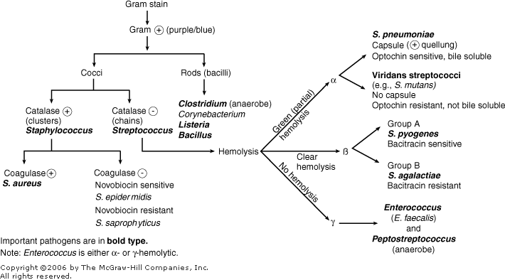 | Lab algorithm for the identification of gram-positive organisms. (Reprinted, with permission, from Bhushan, V [ed.] First Aid for the USMLE, Step 1, McGraw-Hill, 2006.) |
|
| 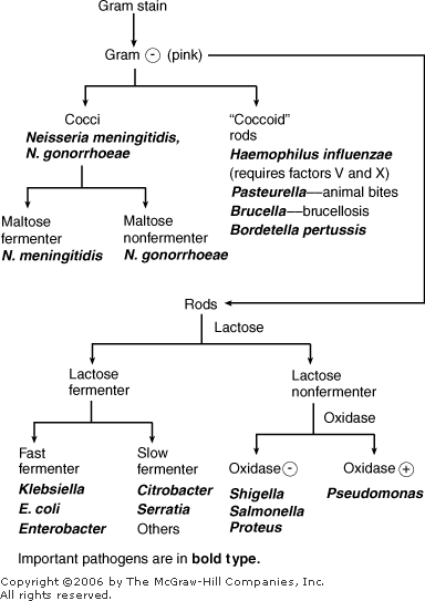 | Lab algorithm for the identification of gram-negative organisms. (Reprinted, with permission, from Bhushan, V [ed.] First Aid for the USMLE, Step 1, McGraw-Hill, 2006.) |
|
Table 7 1 Gram Stain Characteristics and Key Features of Common Organisms
|
| | Gram Staining Pattern and Organisms | Identifying Key Features |
|---|
| Gram-Positive Cocci | | | Enterococcus spp. (E. faecalis) (Note: These are equivalent group D Streptococcus) | Pairs, chains; catalase-negative | | Peptostreptococcus spp. | Anaerobic | | Staphylococcus spp. | Clusters; catalase-positive | | Staphylococcus aureus | Clusters; catalase-positive; coagulase-positive; beta-hemolytic; yellow pigment | | Staphylococcus epidermidis | Clusters; catalase-positive; coagulase-negative; skin flora | | Staphylococcus saprophyticus | Clusters; catalase-positive; coagulase-negative | | Streptococcus spp. | Pairs, chains, catalase-negative | | Streptococcus agalactiae (group B) | Pairs, chains; catalase-negative; vaginal flora | | Streptococcus bovis (group D Enterococcus) | Pairs, chains; catalase-negative | | Streptococcus faecalis (group D Enterococcus) | Pairs, chains; catalase-negative | | Streptococcus pneumoniae (Pneumococcus, group B) | Pairs, lancet-shaped; alpha-hemolytic; optochin-sensitive | | Streptococcus pyogenes (group A) | Beta-hemolytic | | Streptococcus viridans | Pairs, chains; catalase-negative; alpha-hemolytic, optochin-resistant | | Gram-Negative Cocci | | | Acinetobacter spp. | Filamentous, branching pattern | | Moraxella (Branhamella) catarrhalis | Diplococci in pairs | | Neisseria gonorrhoeae (gonococcus) | Diplococci in pairs, often intracellular; ferments glucose but not maltose | | Neisseria meningitidis (meningococcus) | Diplococci in pairs; ferments glucose and maltose | | Veillonella spp. | Anaerobic | | Gram-Positive Bacilli | | | Actinomyces | Branching, beaded, rods; anaerobic | | Bacilli anthracis (anthrax) | Spore-forming rod | | Clostridium spp. (C. difficile, C. botulinum, C. tetani) | Large, with spores; anaerobic | | Corynebacterium spp. (C. Diphtheriae) | Small, pleomorphic diphtheroid; skin flora | | Eubacterium spp. | Anaerobic | | Lactobacillus spp. | Common vaginal bacterium; anaerobic | | Listeria monocytogenes | Beta-hemolytic | | Mycobacterium spp. (limited staining) | Only rapidly growing species gram stain (M. abscessus, M. chelonae, M. fortuitum) | | Nocardia | Beaded, branched rods; partially acid-fast-staining | | Propionibacterium acne | Small, pleomorphic diphtheroid; anaerobic | | Gram-Negative Bacilli | | | Acinetobacter spp. | Lactose-negative, oxidase-negative | | Aeromonas hydrophilia | Lactose-negative (usually) oxidase-positive | | Bacteroides fragilis | Anaerobic | | Bordetella pertussis | Coccoid rod | | Brucella (brucellosis) | Coccoid rod | | Citrobacter spp. | Lactose-positive (usually) | | Enterobacter spp. | Lactose-positive (usually) | | Escherichia coli | Lactose-positive | | Fusobacterium spp. | Long, pointed shape; anaerobic | | Haemophilus ducreyi | Gram-negative bacilli | | Haemophilus influenzae | Coccoid rod, requires chocolate agar to support growth | | Klebsiella spp. | Lactose-positive | | Legionella pneumophila | Stains poorly, use silver stain and special medium | | Morganella morganii | Lactose-negative, oxidase-negative | | Proteus mirabilis | Lactose-negative, oxidase-negative, indole-negative | | Proteus vulgaris | Lactose-negative, oxidase-negative, indole-positive | | Providencia spp. | Lactose-negative, oxidase-negative | | Pseudomonas aeruginosa | Lactose-negative, oxidase-positive blue-green pigment | | Salmonella spp. | Lactose-negative, oxidase-negative | | Serratia spp. | Lactose-negative, oxidase-negative | | Serratia marcenscens | Lactose-negative, oxidase-negative, red pigment | | Shigella spp. | Lactose-negative, oxidase-negative | | Stenotrophomonas (Xanthomonas) maltophilia | Lactose-negative, oxidase-negative | | Vibrio chloerae (cholera) | Gram-negative bacilli | | Yersinia enterocolitica | Gram-negative bacilli | | Yersinia pestis (bubonic plague) | Gram-negative bacilli |
|
aOrganisms are aerobic unless otherwise specified. |
India Ink Preparation India ink is used primarily on CSF to identify fungal organisms (especially cryptococci). KOH Preparation KOH (potassium hydroxide) preps are used to diagnose fungal infections and are often a bedside procedure. Vaginal KOH preps are discussed in Chapter 13, Pelvic Examination. - 1. Apply the specimen (vaginal secretion, sputum, hair, skin scrapings) to a slide. Obtain skin scrapings of a lesion by gentle scraping with a no. 15 scalpel blade (see Figure 13 1 B for description).
- 2. Add 1 or 2 gtt of 10% KOH solution and mix. Gentle heating (optional) may accelerate dissolution of the keratin. A fishy odor from a vaginal prep suggests the presence of Gardnerella vaginalis (see Pelvic Examination).
- 3. Put a coverslip over the specimen, and examine for branching hyphae and blastospores, which indicate the presence of a fungus. KOH should destroy most elements other than fungus. If dense keratin and debris are present, allow the slide to sit for several hours and then repeat the microscopic examination. Lowering the substage condenser provides better contrast between organisms and background.
Lyme Disease Testing Order only when there is real clinical suspicion and not to screen patients with vague, nonspecific complaints but no exposure to the tick vector (Ixodes dammini in the eastern United States and Ixodes pacificus in the western United States). (See also Chapter 4.) The EIA is a screening test and is highly sensitive. If the EIA result is positive or indeterminate, a Western blot is used to confirm or to disprove Lyme disease. The Western blot is performed in two parts: an IgM assay (positive if 2 of 3 bands are positive) and an IgG assay (positive if 5 of 10 bands are positive). Malaria Smear Travelers with fever who have recently returned from regions where malaria is endemic need to have malaria ruled out because certain forms can be rapidly fatal. Prophylaxis is never 100%, and breakthrough infections occur. The reference standard for malaria diagnosis is serial examination of sets of thick and thin blood smears. A single set of smears is never adequate for ruling out malaria. - 1. Perform smears every 12 h until the diagnosis is made or excluded. A rapid dipstick test also can be performed. The thin smears are used to identify the species of malaria, which is used to guide treatment.
- 2. Report confirmed cases of malaria to the CDC. The reporting form is available at http://www.cdc.gov/malaria/clinicians.htm#case (accessed July 5, 2006)
Molecular Microbiology Molecular techniques are used to identify many bacterial and viral organisms without culturing. Many tests rely on DNA probes to identify the pathogens (eg, Gen-Probe). The following microbes are commonly identified from clinical specimens (ie, swab, serum, tissue). Availability varies by clinical facility. Common microorganisms identifiable with a PCR/DNA probe are Chlamydia trachomatis, Borrelia burgdorferi (Lyme disease), HIV, Mycoplasma pneumoniae, M. tuberculosis, N. gonorrhoeae, hepatitis B, and HPV. Other probes are under development. Nasal Culture Obtain the specimen from deep in the nasopharynx and not the anterior nares. Do not let the swab touch the skin. Curved swabs are available through most pathology labs. If resistance is met in one nares, try the other side and do not force. Gently rotate the swab and attempt to rest there for 20 s. Place the swab in the provided tube (eg, Culturette). Cultures of nasopharyngeal specimens are useful in identifying S. aureus and Neisseria meningitidis infections and carriers. Normal nasal flora include S. epidermidis, S. aureus, Streptococcus pneumoniae, Haemophilus influenzae, and several others. Pinworm Preparation (Cellophane Tape Test) Used to identify infestation with Enterobius vermicularis. Wrap a 3-in piece of clear cellophane (eg, Scotch) tape around a glass slide (sticky side out). Touch the slide to the patient's perianal skin in four quadrants and examine it under a microscope to find pinworm eggs. The best sample is collected either in the early morning before bathing or several hours after retiring. Sputum Culture (See Pulmonary Infection) Stool Culture (See Infectious Diarrhea) Stool Leukocyte Stain (See Infectious Diarrhea) Stool for Ova and Parasites If a patient has toxic diarrhea, consider the possibility of parasitic disease, and order a stool for "ova and parasites." Protozoa (Blastocystis, Giardia, and amebae such as Entamoeba histolytica) cannot be cultured and are identified when mature, mobile organisms or cysts are found at microscopic examination of freshly passed feces. Immunosuppressed (eg, HIV-positive) persons may have infection with Cryptosporidium, microsporidia, and Isospora belli. Strongyloides often causes GI symptoms in persons with normal immune function. Ova of parasites such as nematodes (Ascaris, Strongyloides), cestodes (Taenia, Hymenolepis), and trematodes (Schistosoma) are frequently identified in the stool. Syphilis Testing (See Chapter 4) Throat Culture Used to differentiate viral from bacterial (usually group A beta-hemolytic streptococci, eg, Streptococcus pyogenes) pharyngitis. - 1. Obtain the sample with a tongue blade and a good light source.
- 2. Do not attempt a culture if epiglottitis (croup) is suspected (stridor, drooling).
- 3. Use the culture swab and try to touch only the involved area, not the oral mucosa or tongue. If the patient is uncooperative, use an archlike swath that touches both the tonsillar areas and the posterior pharynx.
- 4. If N. gonorrhoeae infection is suspected, use Thayer Martin medium.
- 5. Culture diphtheria (C. diphtheriae, which has a characteristic pseudomembrane) on special medium and notify the lab.
Throat cultures take 24 48 h for completion. An office-based rapid antigen assay also can be performed. Because the sensitivity of this test is only about 80%, the culture provides a "backup" in the case of a false-negative result. Such a small delay in the start of antibiotics does not increase the risk of development of rheumatic fever. Normal flora on routine throat culture can include alpha-hemolytic streptococci, nonhemolytic Staphylococcus, saprophytic Neisseria spp., Haemophilus, Klebsiella, Candida, and diphtheroids. Tzanck Smear Named after Arnault Tzanck. Used in the diagnosis of herpesvirus infections (ie, herpes zoster or simplex). - 1. Clean a cutaneous vesicle (not a pustule or crusted lesion) with alcohol, allow it to air dry, and gently unroof it with a no. 15 scalpel blade.
- 2. Scrape the base of the vesicle with the blade, and place the material on a glass slide. After the specimen air dries, stain the slide with Wright or Giemsa stain. Use high power and oil immersion to identify multinucleated giant cells (epithelial cells infected with herpesviruses). The presence of these cells strongly suggests viral infection; culture is necessary to identify the specific virus.
Urine Culture (See Common Clinical Applications, Urinary Tract Infections [UTI]) Vaginal Wet Preparation (See Chapter 13, Pelvic Examination) Viral Cultures and Serology The laboratory provides the proper collection container for the specific virus. Common pathogenic viruses cultured include herpes simplex (from genital vesicles, throat), CMV (from urine or throat), varicella-zoster (from skin vesicles in children with chickenpox and adults with shingles), and enterovirus (rectal swab, throat). For serologic testing, obtain an acute specimen (titer) as early as possible in the course of the illness, and take a convalescent specimen (titer) 2 4 wk later. A convalescent titer fourfold greater than the acute titer indicates active infection (see Chapter 4 for selected viral antibody titers). With the development of PCR techniques, biopsies performed on older lesions may yield useful information when cultures are negative. |
Differential Diagnosis of Common Infections and Empiric Therapy The pathogens causing common infectious diseases are outlined in Table 7 2 along with empiric therapeutic recommendations. The antimicrobial drug of choice for the management of infection is usually the most active drug against the pathogenic organism or the least toxic alternative. The choice of drugs is modified by the site of infection, clinical status (allergy, renal disease, pregnancy, etc), and susceptibility testing. Table 7 2 Organisms that Cause Common Infectious Diseases with Recommended Empiric Therapya
|
| | Site/Condition | Common Uncommon but Important | Common Empiric Therapy (Modify based on clinical factors such as Gram stain) |
|---|
| BONES AND JOINTS | | | | Osteomyelitis | Staphylococcus aureus | Oxacillin, nafcillin | | Enterobacteriaceae | | If nail puncture: Pseudomonas spp. | | Joint, septic arthritis | S. aureus | Oxacillin; ceftriaxone if gonococci | | Group A strep | | Enterobacteriaceae | | Gonococci | | Joint, prosthetic | S. aureus, S. epididymis, Streptococcus spp. | Vancomycin plus ciprofloxacin | | BREAST | | | | Mastitis, postpartum | S. aureus | Cefazolin, nafcillin, oxacillin | | BRONCHITIS | In adolescent/young patient: Mycoplasma pneumoniae | Treatment controversial because most infections are viral; treat if febrile, associated with sinusitis, positive sputum culture in patients with COPD, or if duration >7 days; doxycycline, erythromycin, azithromycin, clarithromycin | | Respiratory viruses | | In chronic adult infection: Streptococcus pneumoniae, Haemophilus influenzae, Moraxella catarrhalis | | Chlamydia pneumoniae | | CERVICITIS (nongonococcal) | Chlamydia, Mycoplasma hominis, Ureaplasma, others | Azithromycin single dose, doxycycline (evaluate and treat partner) | | CHANCROID | Haemophilus ducreyi | Ceftriaxone or azithromycin as single dose | | CHLAMYDIA | | | | Urethritis, cervicitis, conjunctivitis, proctitis | Chlamydia trachomatis | Azithromycin, doxycycline (amoxicillin if pregnant) | | Neonatal ophthalmia, pneumonia | | Erythromycin | | Lymphogranuloma venereum | C. trachomatis (specific serotypes, L1, L2, L3) | Doxycycline | | DIVERTICULITIS (no perforation or peritonitis) | Enterobacteriaceae, enterococci, bacteroids | TMP SMX, ciprofloxacin plus metronidazole | | EAR | | | | Acute mastoiditis | S. pneumoniae | Amoxicillin, ampicillin/clavulanic acid, cefuroxime | | Group A strep | | S. aureus | | Chronic mastoiditis | Polymicrobial: Anaerobes | Ticarcillin/clavulanic acid, imipenem | | Enterobacteriaceae | | Rarely: Mycobacterium tuberculosis | | Otitis externa | Pseudomonas spp. | Topical agents such as Cortisporin otic, TobraDex | | Enterobacteriaceae | | | In diabetic or malignant otitis: Pseudomonas spp. | Malignant otitis externa: acute aminoglycoside, plus ceftazidime, imipenem, or piperacillin | | Otitis media | S. pneumoniae, H. influenzae, M. catarrhalis, viral causes | Amoxicillin, ampicillin/clavulanic acid, cefuroxime | | S. aureus, group A strep | | In nasal intubation: Enterobacteriaceae, Pseudomonas spp. | | EMPYEMA ENDOCARDITIS | S. pneumoniae, S. aureus | Cefotaxime, ceftriaxone | | Native valve | S. viridans | Parenteral: penicillin or ampicillin or oxacillin or nafcillin plus gentamicin; vancomycin plus gentamicin | | S. pneumoniae | | Enterococci | | S. bovis | | IV drug use | S. aureus | Nafcillin plus gentamicin | | Pseudomonas spp. | | Prosthetic valve | If early (<6 mo after implantation) | Vancomycin plus rifampin plus gentamicin | | S. epidermidis | | S. aureus | | Enterobacteriaceae | | If late (>6 mo after implantation) | | S. viridans | | Enterococci | | S. epidermidis | | S. aureus | | EPIGLOTTITIS | H. influenzae | Ceftriaxone, cefotaxime, cefuroxime ampicillin/sulbactam, or TMP SMX | | S. pneumoniae | | S. aureus | | Group A strep | | GALLBLADDER | | | | Cholecystitis | Acute: E. coli, Klebsiella, Enterococcus | Ampicillin plus gentamicin w/wo metronidazole, imipenem | | Chronic obstruction: anaerobes, coliforms, Clostridium | | Cholangitis | E. coli, Klebsiella, Enterococcus | | | GASTROENTERITIS | | | | Afebrile, no gross blood or WBC in stool | Virus, mild bacterial form | Supportive care only | | Febrile, gross blood, WBC in stool | Enteropathogenic E. coli | Empiric treatment pending cultures: ciprofloxacin, levofloxacin | | Shigella | | Salmonella | | Campylobacter | | Vibrio | | C. difficile | | Listeria monocytogenes | | GRANULOMA INGUINALE GONORRHEA (urethra, cervix, rectum, pharynx) | Calymmatobacterium granulomatis | Doxycycline, TMP SMX | | Neisseria gonorrhoeae | Cefixime, ciprofloxacin, ofloxacin, ceftriaxone all as single dose; treat also for chlamydia | | MENINGITIS (Empiric therapy before cultures) | | | | Neonate | Group B streptococci, E. coli, L. monocytogenes | Ampicillin plus cefotaxime | | Infant 1 3 mo | S. pneumoniae | | N. meningitidis | | Child/adult, community acquired | S. pneumoniae | Vancomycin plus ceftriaxone | | N. meningitidis, H. influenzae | | Postoperative or traumatic | S. epidermitis, S. aureus, S. pneumoniae, Pseudomonas | Vancomycin plus ceftazidime | | Immunosuppressed (ie, steroids) | Gram-negative bacilli, L. monocytogenes | Ampicillin plus ceftazidime | | History of alcohol abuse | S. pneumoniae, L. monocytogenes | Ampicillin plus ceftriaxone or cefotaxime plus vancomycin | | Pseudomonas spp. | | H. influenzae | | HIV infection | Cryptococcus | Amphotericin B (acutely), fluconazole | | NOCARDIOSIS | Nocardia asteroides | Sulfisoxazole, TMP SMX | | PELVIC INFLAMMATORY DISEASE | Gonococci | Ofloxacin and metronidazole or ceftriaxone (single dose) plus doxycycline; parenteral cefotetan or cefoxitin plus doxycycline | | Enterobacteriaceae | | Bacteroides spp. | | Chlamydia | | Enterococci | | M. hominis | | PERITONITIS | | | | Primary (spontaneous) | S. pneumoniae | Cefotaxime or ceftriaxone | | Enterobacteriaceae | | Secondary (to bowel perforation, etc.) | Enterobacteriaceae, Bacteroides spp. | Suspect small bowel: piperacillin, mezlocillin, meropenem, cefoxitin | | Enterococci | Suspect large bowel: clindamycin plus aminoglycosides | | | Pseudomonas spp. | | | Peritoneal, dialysis related | S. epidermidis | Based on culture | | S. aureus | | Enterobacteriaceae | | Candida | | PHARYNGITIS | Respiratory virus | Exudative (group A strep): benzathine penicillin G, erythromycin, loracarbef, azithromycin | | Gonococci | | C. diphtheria | | Epstein Barr virus (infectious mono); spirochetes, anaerobes | | PNEUMONIA | | | | Neonate | Viral (CMV, herpes), bacterial (group B strep, L. monocytogenes, coliforms, S. aureus, Chlamydia) | Ampicillin or nafcillin plus gentamicin | | Infants (1 24 mo) | Most viral such as RSV; S. pneumoniae, Chlamydia, Mycoplasma | Cefuroxime; if critically ill, cefotaxime, ceftriaxone plus cloxacillin | | Child (3 mo 5 y) | As above | Erythromycin, clarithromycin; if critically ill, cefuroxime plus erythromycin | | Child (5 18 y) | Mycoplasma, respiratory viruses, S. pneumoniae, C. pneumoniae | Clarithromycin, azithromycin; erythromycin | | Adult community-acquired | M. pneumoniae, C. pneumoniae, S. pneumoniae | Clarithromycin, azithromycin | | Smokers: as above plus M. catarrhalis, H. influenzae | If hospitalized, third-generation cephalosporin plus azithromycin | | Adult community-acquired aspiration | S. pneumoniae and flora, including anaerobes (eg, Fusobacterium, Bacteroides spp.) | | | Enterobacteriaceae | | Adult community-acquired or ventilator-associated | S. pneumoniae, coliforms, Pseudomonas, Legionella | Imipenem, meropenem | | HIV-associated | Pneumocystis | Pneumocystis: TMP SMX; may require steroids | | Others, as above | | TB, fungi | | SINUSITIS | S. pneumoniae | Acute: TMP SMX ampicillin, amoxicillin/clavulanic acid, clarithromycin | | H. influenzae | | M. catarrhalis | | Anaerobes | | In nosocomial, nasal intubations, etc.: | | S. aureus | | Pseudomonas spp. | | Enterobacteriaceae | | SKIN/SOFT TISSUE | | | | Acne | Propionibacterium acne | Tetracycline, minocycline, topical clindamycin | | Acne rosacea | Possible skin mite | Topical: metronidazole, doxycycline | | Burns | S. aureus, Enterobacteriaceae, Pseudomonas, Proteus | Topical: silver sulfadiazine | | Herpes simplex virus, Providencia, Serratia, Candida | Sepsis: Aztreonam or tobramycin plus cefoperazone, ceftazidime or piperacillin | | Bite (human and animal) | Anaerobes | Ampicillin/sulbactam IV or amoxicillin/clavulanic acid PO | | P. multiloculada | | Cellulitis | Streptococcus spp. (group A, B, C, G) | Diabetic: nafcillin, oxacillin with or without penicillin; if anaerobic, high-dose penicillin G, cefoxitin, cefotetan | | Anaerobic | | Decubitus | Group A strep (S. pyogenes) | If acutely ill: imipenem, meropenem, ticarcillin/clavulanic acid | | Anaerobes, S. aureus, Enterobacteria | | Polymicrobial anaerobic | | Erysipelas | Group A strep (S. pyogenes) | Nafcillin, oxacillin, dicloxacillin, cefazolin | | Impetigo | Group A strep | Penicillin, erythromycin, oxacillin or nafcillin if S. aureus | | S. aureus | | Tinea capitis (scalp) "ringworm" | Fungus: Trichophyton spp., Microsporum spp. | Terbinafine, itraconazole, fluconazole | | Tinea corporis (body) | Fungus: Trichophyton spp., Epidermophyton | Topical: ciclopirox, clotrimazole, econazole, ketoconazole, miconazole, terconazole, others | | Tinea unguium | Various fungi | Itraconazole, fluconazole, terbinafine | | SYPHILIS (less than 1 y duration) | Treponema pallidum | Benzathine penicillin G one dose, doxycycline, tetracycline, ceftriaxone | | TUBERCULOSIS | Mycobacterium tuberculosis | | | Pulmonary, HIV ( ) | | INH, rifampin ethambutol plus pyrazinamide at least 6 mo (+/ pyridoxine) | | TB exposure, PPD ( ) | | Children <5 INH x3 mo (+/ pyridoxine), repeat PPD in 3 mo, others observe | | Prophylaxis in high-risk patients (diabetics, IV drug users, immunosuppressed, etc.) | | INH 6 12 mo (+/ pyroxidine) | | PPD + conversion | | INH 6 12 mo (+/ pyridoxine) | | ULCER DISEASE (duodenal or gastric, not NSAID related) | Helicobacter pylori | Omeprazole plus amoxicillin plus clarithromycin | | URINARY TRACT INFECTION | | | | Cystitis | Enterobacteriaceae (E. coli most common) | Quinolone, TMP SMX | | Staphylococcus saprophyticus (young female) | | Candida | Candida: fluconazole or amphotericin B bladder irrigation | | Urethritis | Gonococci, C. trachomatis, Trichomonas | Ceftriaxone, cefixime, ciprofloxacin, ofloxacin (all one dose) plus azithromycin (single dose) or doxycycline (treat partner) | | Herpesvirus | | Ureaplasma urealyticum | | Prostatitis, acute <35 y | C. trachomatis | Ofloxacin | | Gonococci | | Coliforms | | Cryptococcus (AIDS) | | Prostatitis, acute >35 y | Coliforms | Quinolone, TMP SMX; if acutely ill gentamicin/ampicillin IV | | Prostatitis, chronic bacterial | Coliforms, enterococci, Pseudomonas | Long-term ciprofloxacin or ofloxacin | | Pyelonephritis | Enterobacteriaceae (E. coli) | If acutely ill, gentamicin/ampicillin IV; quinolone, TMP SMX | | Enterococci | | Pseudomonas spp. | | VAGINA | | | | Candidiasis | C. albicans | Fluconazole, itraconazole | | C. glabrata, C. tropicalis | | Trichomonas | Trichomonas vaginalis | Metronidazole (treat partner) | | Vaginosis, bacterial | Polymicrobial (Gardnerella vaginalis, Bacteroides, M. hominis) | Metronidazole (PO or vaginal gel); clindamycin, PO or intravaginally |
|
aAll antimicrobial therapy should be based on complete clinical data, including results of Gram stains and cultures. See also Tables 7 3 (Viral), 7 4 (HIV), 7 5 (Fungal), 7 6 (Parasitic), and 7 7 (Tick-Borne). Note: These guidelines are based on agents commonly involved in adult infections. Actual anti-microbial treatment should be guided by microbiologic studies interpreted in the clinical setting. INH = isoniazid; TMP SMX = trimethoprim sulfamethoxazole. |
Tables 7-2, 7-3, 7-4, 7-5, 7-6, and 7-7 provide empiric treatment guidelines for common infectious diseases, including bacterial, viral, HIV, fungal, parasitic, and tick-borne diseases. Additional resources include The Sanford Guide to Antimicrobial Therapy (www.sanfordguide.com) and the Johns Hopkins ABX Guide (www.hopkins-abxguide.org). Table 7 3 Pathogens and Drugs of Choice for Treating Common Viral Infectionsa
|
| | Viral Infection | Drug of Choice | Adult Dosage |
|---|
| CMV | | | | Retinitis, colitis, esophagitis | Ganciclovir (Cytovene)b
| 5 mg/kg IV q12h x 14 21d, 5 mg/kg/d IV or 6 mg/kg IV 5x/wk or 1 g PO tid | (Vitrasertb) implants or foscarnet (Foscavir)
| 4.5 mg intraocularly q 5 8 mo | | | 60 mg/kg IV q8h or 90 mg/kg IV q1 2 h x 14 21 d followed by 90 120 mg/kg/d IV | | or cidofovir (Vistide) | 5 mg/kg/wk IV x 2 wk, then 5 mg/kg IV q2 wk | | or fomivirsen (Vitravene) | 330 mcg intravitreally q2 wk x 2 then 1/mo | | EBV | | | | Infectious mononucleosis | None | | | HAV | None, but gamma globulin within 2 wk of exposure may limit infection | 0.2 mL/kg IM x 1 | | HBV | | | | Chronic hepatitis | Lamivudine (Epivir HBV) | 100 mg PO 1x/d x 1 3 y | | Interferon alfa-2b (Intron A) | 5 million units/d or 10 million units 3x/wk SC or IM x 4 mo | | HCV | | | | Chronic hepatitis | Interferon alfa-2b plus ribavirin (Rebetron) | 3 million units 3x/wk SC plus ribavirin 1000 1200 mg/d PO x 2 mo | | Interferon alfa-2b (Intron A) | 3 million units SC or IM 3x/wk x 12 24 mo | | Interferon alfa-2a (Roferon-A) | 3 million units SC or IM 3x/wk x 12 24 mo | | Interferon alfacon-1 (Infergen) | 9 mcg 3x/wk x 6 mo | | HIV (See Table 7 4) | | | | HSV | | | | Orolabial herpes in the immunocompetent with multiple recurrences | Penciclovir (Denavir) | 1% cream applied q2h while awake x 4 d | | Genital herpes | | | | First occurrence | Acyclovir (Zovirax) | 400 mg PO tid or 200 mg PO 5x/d x 7 10 d | | or famciclovir (Famvir) | 250 mg PO tid x 5 10 d | | or valacyclovir (Valtrex) | 1 g PO bid x 7 10 d | | Recurrence | Acyclovir (Zovirax) | 400 mg PO tid x 5 d | | or famciclovir (Famvir) | 125 mg PO bid x 5 d | | or valacyclovir (Valtrex) | 500 mg PO bid x 5 d | | Chronic suppression | Acyclovir (Zovirax) | 400 mg PO bid | | or famciclovir (Famvir) | 500 1000 mg PO 1x/d | | or valacyclovir (Valtrex) | 250 mg PO bid | | Mucocutaneous in the immunocompromised | Acyclovir (Zovirax) | 5 mg/kg IV q8h x 7 14 d | | or acyclovir (Zovirax) | 400 mg PO 5x/d x 7 14 d | | Encephalitis | Acyclovir (Zovirax) | 10 15 mg/kg IV q8h x 14 21 d | | Neonatal | Acyclovir (Zovirax) | 20 mg/kg IV q8h x 14 21 d | | Acyclovir-resistant | Foscarnet (Foscavir) | 40 mg/kg IV q8h x 14 21 d | | Keratoconjunctivitis | Trifluridine (Viroptic) | 1 drop 1% solution topically, q2h, up to 9 gtt/d x 10 d | | INFLUENZA A VIRUS | Rimantadine (Flumadine) | 200 mg PO 1x/d or 100 mg PO bid x 5 d | | Amantadine (Symmetrel) | 100 mg PO bid x 5 d | | INFLUENZA A AND B VIRUS | Zanamivir (Relenza) | 10 mg bid x 5d by inhaler | | Oseltamivir (Tamiflu) | 75 mg PO bid x 5 d | | MEASLES | | | | Children | None (immunize, See Chapter 22) | | | Adults | None or ribavirin | 20 35 mg/kg/d x 7 d | | PAPILLOMA VIRUS (HPV) | | | | Anogenital warts | Podofilox or podophyllin | Topical application (see Chapter 22) | | Interferon alfa-2b (Intron A) | 1 million units intralesional 3x/wk x 3 wk | | Imiquimod, 5% cream (Aldara) | Apply 3/wk h, remove 6 10 h later up to 16 wk | | RSV | | | | Bronchiolitis | Ribavirin (Virazole) | Aerosol treatment 1218 h/d x 3 7 d | | VZV | | | | Exposure prophylaxis in the immunocompromised (HIV, steroids, etc.) | Varicella zoster immune globulin | See package insert | | Varicella (>12 y old) | Acyclovir (Zovirax) | 20 mg/kg (800 mg max) PO qid x 5 d | | Herpes zoster | Valacyclovir (Valtrex) | 1 g PO tid x 7 d | | or famciclovir (Famvir) | 500 mg PO tid x 7 d | | or acyclovir (Zovirax) | 800 mg PO 5x/d x 7 10 d | | Varicella or zoster in the immunocompromised | Acyclovir (Zovirax) | 10 mg/kg IV q8h x 7 d | | Acyclovir-resistant | Foscarnet (Foscavir) | 40 mg/kg IV q8h x 10 d |
|
aBased on Guidelines from the CDC published in MMWR and the Medical Letter Vol. 41 December 3, 1999. bThe generic drug name appears in regular type; the trade name appears in parentheses afterward in italics. CMV = cytomegalovirus; EBV = Epstein Barr virus; HAV = hepatitis A virus; HBV = hepatitis B virus; HCV = hepatitis C virus; HIV = human immunodeficiency virus; HPV = human papilloma virus; HSV = herpes simplex virus; RSV = respiratory syncytial virus; VZV = varicella-zoster virus. |
Table 7 4 Antiretroviral Regimens Recommended for Management of HIV-1 Infection in Antiretroviral-Na ve Patients
|
| | Regimen | Drugs | No. of Pills |
|---|
| Preferred Regimens | | | | NNRTI-based | Efavirenz + (lamivudine or emtricitabine) + (zidovudine or tenofovir DF) (AII) (note: efavirenz is not recommended for use in the first trimester of pregnancy or in women with high pregnancy potentiala)
| 2 3 | | PI-based | Lopinavir/ritonavir (coformulation) + (lamivudine or emtricitabine) + zidovudine (AII) | 6 7 | | Alternative Regimens | | | | NNRTI-based | Efavirenz + (lamivudine or emtricitabine) + (abacavir or didanosine or stavudine) (BII) (note: efavirenz is not recommended for use in the first trimester of pregnancy or in women with high pregnancy potentiala)
| 2 4 | | | Nevirapine + (lamivudine or emtricitabine) + (zidovudine or stavudine or didanosine or abacavir or tenofovir) (BII) [note: high incidence (11%) of symptomatic hepatic events was observed in women with pre-nevirapine CD4 T cell counts >240 cells/mm3 and men with CD4 T cell counts >400 cells/mm3 (6.3%). Nevirapine should not be initiated in these patients unless the benefit clearly outweighs the risk.]
| 3 6 | | PI-based | Atazanavir + (lamivudine or emtricitabine) + (zidovudine or stavudine or abacavir or didanosine) or (tenofovir + ritonavir 100 mg/d) (BII) | 3 6 | | | Fosamprenavir (lamivudine or emtricitabine) + (zidovudine or stavudine or abacavir or tenofovir or didanosine) (BII) | 5 8 | | | Fosamprenovir/ritonavirb + (lamivudine or emtricitabine) + (zidovudine or stavudine or abacavir or tenofovir or didanosine) (BII)
| 5 8 | | | Indinavir/ritonavirb + (lamivudine or emtricitabine) + (zidovudine or stavudine or abacavir or tenofovir or didanosine) (BII)
| 7 12 | | | Lopinavir/ritonavir + (lamivudine or emtricitabine) + (stavudine or abacavir or tenofovir or didanosine) (BII) | 5 8 | | | Nelfinavir + (lamivudine or emtricitabine) + (zidovudine or stavudine or abacavir or tenofovir or didanosine) (CII) | 5 8 | | | Saquinavir (sgc, hgc, or tablets)c/ritonavirb + (lamivudine or emtricitabine) + (zidovudine or stavudine or abacavir or tenofovir or didanosine) (BII)
| 7 15 | | 3 NRTI-based | Abacavir + zidovudine + lamivudine only when a preferred or an alternative NNRTI- or PI-based regimen cannot or should not be used (CII) | 2 |
|
aWomen with child-bearing potential implies women who want to conceive or those who are not using effective contraception. bLow-dose (100 400 mg) ritonavir per day. csgc = soft gel capsule; hgc = hard gel capsule. AII = Strong recommendation with the clinical trial results; BII = Moderate recommendation with clinical trial results; CII = Optional recommendation with clinical trials results; NRTI = two nucleoside/nucleotide reverse transcriptase inhibitors; NNRTI = either a nonnucleoside reverse transcriptase inhibitor or a ritonavir-boosted or unboosted protease inhibitor; PI = protease inhibitor. http://aidsinfo.nih.gov/contentfiles/adultandadolescentGL.pdf (accessed July 3, 2006) |
Table 7 5 Systemic Drugs for Managing Fungal Infections
|
| | Infection | Drug of Choice | Alternatives |
|---|
| ASPERGILLOSIS | Amphotericin B or itraconazole | Amphotericin B lipid complex, amphotericin cholesteryl complex liposomal amphotericin B | | BLASTOMYCOSIS | Itraconazole or amphotericin B | Fluconazole | | CANDIDIASIS | | | | Oral (thrush) | Fluconazole or itraconazole | Nystatin lozenge or swish and swallow | | Stomatitis, esophagitis, vaginitis in AIDS | Fluconazole or itraconazole | Parenteral or oral amphotericin B | | Systemic | Amphotericin B or fluconazole | | | Cystitis/vaginitis | See Table 7 2 | | | COCCIDIOIDOMYCOSIS | | | | Pulmonary (otherwise healthy) | No drug usually recommended | | | Pulmonary (high risk) | Itraconazole or fluconazole | Amphotericin B | | CRYPTOCOCCOSIS | | | | In non-AIDS patient | Amphotericin B or fluconazole | Amphotericin B fluconazole | | Meningitis (HIV/AIDS) | Amphotericin B plus 5-flucytosine; then long-term suppression with fluconazole | Amphotericin B lipid complex | | HISTOPLASMOSIS | | | | Pulmonary, disseminated | | | | Otherwise healthy | Moderate disease: itraconazole | Severe: amphotericin B | | HIV/AIDS | Amphotericin B, followed by itraconazole suppression | Itraconazole | | MUCORMYCOSIS | Amphotericin B | No dependable alternative | | PARACOCCIDIOIDOMYCOSIS | Itraconazole | Amphotericin B | | SPOROTRICHOSIS | | | | Cutaneous | Itraconazole | Potassium iodide 1 5 mL tid | | Systemic | Itraconazole | Amphotericin B |
|
|
Table 7 6 Drugs for Treating Selected Parasitic Infections
|
| | Infection | Drug |
|---|
| Amebiasis (Entamoeba histolytica) | | | Asymptomatic | Iodoquinol or paramomycin | | Mild to moderate intestinal disease | Metronidazole or tinidazole | | Severe intestinal disease, hepatic abscess | Metronidazole or tinidazole | | Ascariasis (Ascaris lumbricoides, roundworm) | Albendazole, mebendazole or pyrantel pamoate | | Cryptosporidiosis (Cryptosporidium) | Paromomycin | | Cutaneous larva migrans (creeping eruption, dog and cat hookworm) | Albendazole, thiabendazole or ivermectin | | Cyclospora infection | Trimethoprim sulfamethoxazole | | Enterobius vermicularis (pinworm) | Pyrantel pamoate, mebendazole or albendazole | | Filariasis (Wuchereria bancrofti, Brugia malayi, Loa loa) | Diethylcarbamazine | | Giardiasis (Giardia lamblia) | Metronidazole | | Hookworm infection (Ancylostoma duodenale, Necator americanus) | Albendazole, mebendazole, or pyrantel pamoate | | Isosporiasis (Isospora belli) | Trimethoprim sulfamethoxazole | | Lice (Pediculus humanus, P. capitis, Phthirus pubis) | 1% permethrin (topical) or 0.5% malathion | | Malaria (Plasmodium falciparum, P. ovale, P. vivax, and P. malariae) | | | Chloroquine-resistant P. falciparum | Quinine sulfate plus doxycycline, tetracycline, clindamycin or pyrimethamine sulfadoxine (oral) | | Chloroquine-resistant P. vivax | Quinine sulfate plus doxycycline, or pyrimethamine sulfadoxine (oral) | | All Plasmodium except chloroquine-resistant P. falciparum | Chloroquine phosphate (oral) | | All Plasmodium (parenteral) | Quinine gluconate or quinine dihydrochloride | | Prevention of relapses: P. vivax, and P. ovale only | Primaquine phosphate | | Malaria, prevention | | | Chloroquine-sensitive areas | Chloroquine phosphate | | Chloroquine-resistant areas | Mefloquine or doxycycline | | Mites, see Scabies | | | Pinworm, see Enterobius | | | Pneumocystis carinii pneumonia | Trimethoprim sulfamethoxazole | | Alternative: TMP-dapsone, clindamycin-primaquine, pentamidine; mild disease oral, moderate to severe IV plus steroids | | Primary and secondary prophylaxis | Trimethoprim sulfamethoxazole | | Roundworm, see Ascariasis | | | Scabies (Sarcoptes scabiei) | 5% Permethrin topically | | Alternatives: ivermectin, 10% crotamiton | | Strongyloidiasis (Strongyloides stercoralis) | Ivermectin | | Tapeworm infection | | | Adult (intestinal stage) | | | Diphyllobothrium latum (fish), Taenia saginata (beef), Taenia solium (pork), Dipylidium caninum (dog), Hymenolepis nana (dwarf tapeworm) | Praziquantel | | Larval (tissue stage) | | | Echinococcus granulosus (hydatid cyst) | Albendazole | | Cysticercus cellulosae (cysticercosis) | Albendazole or praziquantel | | Toxoplasmosis (Toxoplasma gondii) | Pyrimethamine plus sulfadiazine | | Trichinosis (Trichinella spiralis) | Steroids for severe symptoms plus mebendazole | | Trichomoniasis (Trichomonas vaginalis) | Metronidazole or tinidazole | | Hairworm infection (Trichostrongylus colubriformis) | Pyrantel pamoate | | Trypanosomiasis (Trypanosoma cruzi, Chagas disease) | Benznidazole | | Trichuriasis (Trichuris trichiuria, whipworm) | Mebendazole or albendazole | | Visceral larva migrans, toxocariasis (Toxocara canis) | Albendazole or mebendazole |
|
|
Table 7 7 Guide to Common Tick-Borne Diseases
|
| | | Rocky Mountain Spotted Fever | Human Granulocytic Ehrlichiosis | Lyme Disease | Babesiosis |
|---|
| Causative Agent | Rickettsia rickettsii (bacterium) | Ehrlichia spp. (bacterium) | Borrelia burgdorferi (bacterium) | Babesia microti (protozoan) | | Season | Mostly spring, summer | Peaks in summer, may be seen year-round | Mostly spring, but year-round | Mostly spring/summer | | Vector Habits | American Dog Tick | Deer Tick (black-legged) | Same as for the deer tick | Same as for the deer tick | | | Found in high grass and low shrubs, fields | Found in woodlands, old fields, landscaping with significant ground cover vegetation | | | | | Lone Star Tick | | | | | | Found in woodlands, forest edge, and old fields | | | | | Classical Clinical Presentation | Sudden moderate to high fever, severe headache, maculopapular rash (with planer/palmer presentation) | Fever, headache, constitutional symptoms | EM rash, constitutional symptoms, arthritis, cardiovascular and nervous system involvement | Fever, hemolytic anemia, constitutional symptoms | | Incubation Period | 2 14 d | 1 30 d | 3 30 d | 1 52 wk | | Diagnosis | Clinical serology | Clinical serology | Clinical serology, culture | Thick and thin blood smears | | Treatment | Adults doxycycline | Adults tetracyclines | Doxycycline, amoxicillin, cefuroxime for 14 21 d | Clindamycin/quinine | | | Children/pregnant women chloramphenicol | Children/pregnant women consult specialist | | |
|
EM = erythema multiforme. |
|
Common Clinical Applications Urinary Tract Infection (UTI) Sample Collection - A "midstream" or "clean catch" urine sample (see Chapter 13) gives about 85% accurate results for women and girls and uncircumcised men and boys but can be contaminated, as are specimens from long-term Foley catheters.
- The best urine is obtained through a recently placed Foley catheter or from a "straight cath" to minimize contamination (see Chapter 13). Suprapubic needle aspiration is the most accurate method of obtaining urine but is almost never done in adults because of risks. Any growth in urine obtained with these in-and-out techniques is considered positive.
- Never collect urine directly from a urostomy bag; it will be grossly contaminated. The presence of a urostomy (ie, ileal conduit) necessitates that a catheter be placed to collect the specimen.
- If a urine specimen cannot be taken to the lab within 60 min, refrigerate it.
- Differentiating a true urinary tract infection from contamination or simple colonization can be challenging. Pyuria plus bacteriuria usually equals UTI. In general, if an infection is present, inflammatory cells (except in neutropenia) should be detectable in the urine. UA is used to detect inflammatory WBCs and should be ordered with the culture. The presence of epithelial cells suggests contamination; urinary diversion with a bowel segment is an exception.
Urinalysis: (See also Chapter 6) The leukocyte esterase test is used to detect the granules in neutrophils (not lymphocytes) and is a marker for pyuria. The number of WBCs found is probably the most dependable marker for true pyuria. Enterobacteriaceae (gram-negative rods such as Escherichia coli, Klebsiella, and Proteus) reduce nitrate to nitrite. If the urine nitrite test is positive, > 10,000 gram-negative rods are likely present. Gram-positive cocci do not convert nitrate to nitrite, so a negative nitrite test does not help rule out UTI. Gram stain is not routinely done on urine samples but should be performed if the patient is acutely ill and septic. Identification of either gram-positive cocci or gram-negative cocci can be used to guide initial antibiotic therapy. If the urine Gram stain reveals cocci in chains, infection with an Enterococcus species is likely. Urine Culture: A colony count < 10,000 is insignificant, as is the presence of mixed organisms. A colony count > 100,000 is indicative of true infection. Counts of 10,000 100,000 must be interpreted, usually in conjunction with the UA results and the clinical situation. The presence of more than three organisms usually indicates contamination unless the process is chronic, a GI fistula is present, or the patient has undergone certain types of urinary diversion. Routine cultures are insufficient for detection of N. gonorrhoeae and Chlamydia. Pulmonary Infection Sputum Culture and Stain: An early morning sample is preferred because such samples are more likely to be from the lower airway. A Gram stain can be used to guide therapy; if bacteria can be seen on the Gram stain, they must number > 10,000 and are probably significant. As with Gram stain of any clinical specimen, a high neutrophil to epithelial cell ratio suggests a less contaminated specimen. The presence of > 25 epithelial cells/hpf suggests that the specimen is more spit than sputum and is useless. Steps to improve the quality of the sputum collection include: - 1. Careful instructions to the patient to produce a deep sample. If the patient cannot mobilize the secretions, P&PD along with nebulizer treatments may help, as may nasotracheal suctioning with a specimen trap.
- 2. Most labs do not accept anaerobic sputum cultures (critical in the diagnosis of aspiration pneumonia and lung abscesses) unless obtained by transtracheal aspiration or endobronchial endoscopic collection and submitted in special anaerobic transport media.
- 3. PCP is diagnosed with expectorated sputum about 10% of the time. Therefore a technique such as open lung biopsy or endobronchial lavage must be used. Specialized stains for identifying Pneumocystis carinii include methenamine silver, Giemsa, and toluidine blue.
Pulmonary Viral Culture: Viral cultures are performed on both nasopharyngeal washes and bronchoscopy specimens. The viral culture medium must be refrigerated. After the specimen is collected, the medium should be transported to the microbiology lab without delay. Specific cultures should be ordered on the basis of the clinical situation. Examples include herpes simplex virus, varicella-zoster virus, CMV, and other viruses such as enteroviruses. Rapid Influenza Test: Use a nasal swab to detect both influenza A and influenza B; useful when influenza is circulating in the community. Pulmonary TB - 1. Place all patients with possible pulmonary TB in respiratory isolation.
- 2. Evaluate patients with suspected TB with serial sputum acid-fast bacterial smears and culture. Obtain three sputum samples on three separate days. Early morning sputum collection is preferred because the specimen is more likely to be from the lower airway. Prepare an AFB smear to detect acid-fast bacilli (see Microbiology Techniques). If the smear is positive, continue respiratory isolation. Monitor culture results; forward positive cultures to the reference lab for antimycobacterial susceptibility testing.
- 3. If a patient with possible TB is immunosuppressed and the clinical condition is worsening, consider early bronchoscopy and chest CT to better evaluate the lung parenchyma.
- 4. Verify that the mycobacteria recovered are in the M. tuberculosis complex. Techniques are as follows:
- Culture on special growth media, nucleic acid probes, and nucleic acid amplification methods such as PCR. Send all isolates for drug susceptibility testing so proper therapy can be given.
- Because skin testing for TB may not be helpful, order a blood test (T-SPOT.TB or QunatiFERON-TB Gold) to detect serum levels of gamma-interferon in response to specific TB antigens.
- Promptly report confirmed cases of TB to the local public health authorities so that antituberculosis medications can be prescribed. The public health department can also set up direct observed therapy (DOT) to ensure compliance with treatment.
CNS Infection (See also Lumbar Puncture, Chapter 13 ) - 1. Obtain a head CT before LP if any of the following is true: focal neurologic signs are present, a seizure has occurred, optic papilledema is present, the patient's age > 60 y, HIV-1 positive or immunocompromised state is present, a change in mental status has occurred.
- 2. Measure opening pressure. A high opening pressure (> 25 cm water) suggests possible brain herniation and risk of death.
- 3. Order routine tests: cell count, protein, glucose, Gram stain, and bacterial culture.
- 4. Order PCR for the following viruses: herpes simplex, varicella-zoster, and West Nile. Order viral culture for CMV and enterovirus.
- 5. Order a CSF cryptococcal antigen test and an India ink stain to directly visualize the cryptococcal organism.
- 6. Order AFB smear and culture, syphilis testing by VDRL, and a fungal smear and culture if the patient has identifying risk factors on H&P and when sufficient clinical suspicion exists.
- 7. Because cryptococcal meningitis can be life threatening in HIV/AIDS patients, obtain CSF opening pressure because cryptococci can impede the flow of CSF with severe consequences (severe headache, cranial nerve deficits, seizure disorder, and brainstem herniation).
Skin and Soft Tissue Infection The spectrum of skin and soft-tissue infections runs the gamut from routine uncomplicated infections such as cellulitis of an extremity to life-threatening emergencies such as necrotizing fasciitis. Two basic questions need to be addressed. - 1. Is there a collection (eg, abscess) in the region of the infection that has to be drained? If doubt exists, imaging of the infected area can depict pockets of infection and provide guidance on drainage. Such drainage procedures yield material for Gram stain, culture, and sensitivity testing. Fungal staining with culture and acid-fast smears with culture should also be ordered.
- 2. Are contiguous structures already involved or at risk if the infection spreads? Examples of contiguous spread are orbital cellulitis leading to CNS infection and diabetic foot ulcer or sacral decubitus ulcer causing osteomyelitis.
Infectious Diarrhea Stool Culture: A fresh stool sample is cultured for diagnosis of the cause of diarrhea and identification of disease carriers. Most common pathogens Salmonella species, Shigella, and E. coli 0.157 can be grown on standard media. Yersinia and Campylobacter require a special culture medium. Stool Leukocyte Stain (Fecal Leukocytes, L ffler Methylene Blue Stain): The presence of WBCs signifies inflammation in the bowel. Stain differentiates treatable diarrhea (ie, bacterial) from other causes. Used to detect causes of Crohn disease, ulcerative colitis, TB, and amebic infection as well, but many causes of severe diarrhea are viral. The positive predictive value of a bacterial pathogen as a cause of diarrhea is 70%. Clostridia Difficile: Clinical course can be quite variable, ranging from simple diarrhea of short duration to cases of fulminant pseudomembranous colitis, which can be rapidly fatal. The classic triad of fever, leukocytosis, and diarrhea always suggests the possibility of C. difficile colitis. Risk factors for acquisition include antibiotic exposure (clindamycin and cephalosporins most common), certain chemotherapies, advanced age, increasing severity of illness, and anything that disturbs bowel motility, including surgery and medicines. Most labs use ELISA to test for the presence of organisms by detecting antigen (see Chapter 4). The organism must be present if the antigen is present. Two toxins, designated A and B, are routinely sought. Stool for Ova and Parasites: (See Stool for Ova and Parasites) HIV-1 Testing and Screening (See also Chapter 4) The bases of routine HIV-1 testing are the EIA screening test, which is used to detect antibody to the HIV virus, and the confirmatory Western blot. If the screening test for HIV is positive, Western blot testing is performed to confirm true infection. HIV antibodies usually appear 4 16 wk after initial infection occurs. The ability to detect the antibody in the serum of a patient is called seroconversion. Testing for HIV-1 in Acute Retroviral Syndrome: New infection with HIV often manifests as a protean mononucleosis-like illness that has been designated acute retroviral syndrome (estimated incidence 20 90% in different series). The syndrome occurs 1 6 wk after infection and is marked by fever, lymphadenopathy, myalgia, and pharyngitis (nonexudative, unlike the pharyngitis that occurs with mononucleosis). Maculopapular viral exanthema, usually on the trunk, occurs in about 50% of patients. The most important factor in the diagnosis of acute retroviral syndrome is to have a high index of clinical suspicion. Because of its protean nature, the diagnosis is missed in many cases. Testing for acute retroviral syndrome and new asymptomatic HIV-1 infection can be problematic. HIV-1 seroconversion (when specific antibodies become detectable in the serum and can be used for diagnostic purposes) usually occurs 4 16 wk after new infection. HIV-1 P24 antigen appears in the serum usually in the first 2 wk of new infection and remains detectable until the host generates sufficient anti-P24 antibody to neutralize the antigen. The P24 assay has been replaced by plasma HIV-1 RNA PCR viral detection assays. A high viral load (> 100,000 viral copies/mL) in the appropriate clinical setting with an exposure history is highly specific and sensitive for the diagnosis of a new HIV infection. The viral load is almost always > 100,000 copies/mL and often > 1 million copies/mL. A low viral load (< 50,000 viral copies/mL) is probably a false-positive result. Because of its extremely high sensitivity, PCR testing is susceptible to lab contamination. HIV-1 and Pregnancy: Decreasing the incidence of vertical transmission of HIV-1 from mother to infant is an issue of paramount importance. The use of highly active antiretroviral therapy (HAART) in conjunction with scheduled cesarean section and abstinence from breast feeding has led to a drop in vertical transmission rates from 25% to < 2%. The decision to initiate HAART in a pregnant woman is a complex decision requiring discussion and careful assessment of risk to both mother and fetus. Administer HAART to a pregnant woman if she meets the standard criteria for starting HAART, such as a CD4 T-cell count of 200 350 CD4 T cells/mL, a high viral load (> 55,000 viral copies/mL), and the presence of an AIDS-defining illness. Other, more specific criteria include a viral load greater than 1000 copies/mL. Some infectious disease specialists recommend delaying HAART until the 10th 12th week of pregnancy to avoid the early organogenesis period of gestation. Consider resistance testing to detect viral resistance. Baseline Tests after New Diagnosis of HIV and Referral to HIV Specialist: The tests used for most of the major clinical decisions regarding HIV-1 therapy are HIV-1 RNA viral load and CD4 T-cell count. The results are used in decisions about when to initiate HAART, when to change HAART in the face of virologic failure (viral load no longer suppressed), and when to give prophylaxis to prevent opportunistic infections such as PCP, cerebral toxoplasmosis, and disseminated M. avium-intracellulare infection. - 1. Order a series of routine tests such as a comprehensive metabolic panel and a CBC to assess the patient's basic organ function.
- 2. Order a fasting lipid panel because of the risk of adverse lipid effects during treatment with certain classes of HAART, such as protease inhibitors.
- 3. If a patient has an allergy to TMP SMX and will need prophylaxis or develops an allergy during prophylaxis and may be given dapsone, order a G6PD assay to rule out G6PD deficiency, especially if the patient is of Mediterranean or African origin or descent.
- 4. Review the initial treatment in Table 7 4. HIV guidelines are available online at: www.aidsinfo.nih.gov/guidelines
Hepatitis (See also Chapter 4, Hepatitis Testing) Hepatitis A Testing: Acute hepatitis A is diagnosed when anti hepatitis A IgM antibodies are present. These antibodies appear soon after the initial infection and persist for approximately 4 mo. Anti hepatitis A IgG titer increases slowly, reaching its peak approximately 4 mo after infection, and then persists for many years. Hepatitis B Testing: The incubation period of hepatitis B is about 12 wk. Hepatitis B surface antigen (HbsAg) usually appears in the first 10 wk after exposure. If HbsAg remains elevated much beyond the first 6 mo, then the diagnosis of chronic hepatitis B is made. Hepatitis C Testing: HCV screening for both surveillance of the blood supply and routine clinical screening is by detection of anti-HCV antibody. The anti-HCV EIA, however, does not show whether the patient has had previous infection and cleared it (about 15% of patients) or if the patient has active infection. Because the EIA was designed with high sensitivity to screen for disease (ie, to protect the blood supply) false-positive tests are not uncommon. False-positive results are most common in screening of low-risk patients. It is extremely important to realize that anti-HCV antibody does not appear in a newly infected patient until at least 8 10 wk after the infective event. The recombinant immunoblot assay (RIBA) was designed to help stratify the risk of true infection. RIBA results are categorized as negative (no antigen detected), indeterminate (one antigen detected), or positive (two or more antigens detected). The clinical utility of RIBA is somewhat limited. If the EIA is positive in a low-risk patient and the RT-PCR for HCV RNA is negative, a negative RIBA makes the possibility of infection extremely remote. A better approach may be to repeat the HCV RNA RT-PCR and to test aminotransferases in 6 mo. Confirmatory testing is best performed with HCV RT-PCR of clinical specimens. HCV RNA testing is broken down into two categories: - Qualitative testing, which has a lower threshold for detecting virus and is useful for confirming infection.
- Quantification of the viral load, which is more appropriate for monitoring response to therapy.
Genotyping of HCV is performed if treatment is being considered, because some specific genotypes of the virus are more susceptible to therapy than are others. Most of the HCV-infected patients in North America have genotype 1, which is much less likely to clear with the standard intense treatment with ribavirin and pegylated interferon, which has to be given for 48 wk. |
Bioterrorism Recognizing unusual patterns in disease presentation, detecting such events, and having a high clinical index of suspicion are critical. Certain situations or patterns should prompt contacting public health officials or the CDC. Examples are: - Any disease or suspected disease on the Category A, B or C list:
- Category A: Anthrax, smallpox, botulism, viral hemorrhagic fever, tularemia, plague
- Category B (lower mortality than A): Q fever, typhus, melioidosis (or glanders), psittacosis, brucellosis, spread of toxins (eg, Ricin)
- Category C (emerging pathogens with bioweapon potential): multiple-drug-resistant TB, Nipah virus, others.
- A case of disease or cluster of cases in the wrong season, such as an outbreak of "influenza" that occurs in the summer.
- Appearance of an infectious disease in the wrong region, such as a case of coccidioidomycosis in the northeastern United States.
- Fulminant progression of a usually benign infection in a healthy host.
- An infectious disease occurring without the presence of its mandatory vector.
Contact information and additional resources: - CCDC, www.bt.cdc.gov
- CDC Emergency Response Hotline (24/7) (770) 488-7100
- CDC Botulism Hotline (404) 639-2206; after hours (404) 639-2206
|
Subacute Bacterial Endocarditis Prophylaxis The most recent SBE guidelines were released by the American Heart Association in 1997 (JAMA 1997;277:1794) and also in 2005 (Med Lett 2005;47:59). The guidelines specify which patients are at high, moderate, or low risk of bacteremia and indicate which procedures are more likely to be associated with bacterial endocarditis. SBE prophylaxis is recommended only for patients who are at high or moderate risk. See Tables 7 8 and 7 9 for regimens. Table 7 8 SBE Prophylaxis for Oral, Respiratory, or Esophageal Proceduresa
|
| | Prophylaxis | Agent | Regimenb
|
|---|
| Standard prophylaxis | Amoxicillin | Adults: 2 g; children: 50 mg/kg PO 1 h before procedure | | Unable to take oral medications | Ampicillin | Adults: 2 g IM or IV; children: 50 mg/kg or IV 30 min before procedure | | Allergic to penicillin | Clindamycin or | Adults: 600 mg; children: 20 mg/kg PO 1 h before procedure | | Cephalexin or cefadroxil | Adults: 2 g; children; 50 mg/kg PO 1 h before procedure | | Azithromycin or clarithromycin | Adults: 500 mg; children: 15 mg/kg PO 1 h before procedure | | | Adults: 600 mg; children: 20 mg/kg IV 30 min before procedure | | Penicillin allergic and unable to take oral medications | Clindamycin or cefazolin | Adults: 600 mg or 1 g; children: 25 mg/kg IM or IV 30 min before procedure |
|
aSee Subacute Bacterial Endocarditis Prophylaxis for recommended risk groups. bTotal children's dose should not exceed adult dose. |
Table 7 9 SBE Prophylaxis for GU/GI (Excluding Esophageal) Proceduresa
|
| | Patient | Agents | Regimen |
|---|
| High-risk | Ampicillin + gentamicin | Adults: ampicillin 2 g IM/IV + gentamicin 1.5 mg/kg (max 120 mg) within 30 min of procedure; 6 h later, ampicillin 1 g IM/IV or amoxicillin 1 g PO | | Children: ampicillin 50 mg/ kg IM or IV (2 g max) + gentamicin 1.5 mg/kg within 30 min of procedure; 6 h later, ampicillin 25 mg/kg IM/IV or amoxicillin 25 mg/kg PO | | High-risk allergic to ampicillin/amoxicillin | Vancomycin + gentamicin | Adults: vancomycin 1 g IV over 1 2 h + gentamicin 1.5 mg/kg IV/IM (120 mg max); dose within 30 min of starting procedure | | Children: vancomycin 20 mg/kg IV over 1 2 h + gentamicin 1.5 mg/kg IV/IM; complete dose within 30 min of starting procedure | | Moderate-risk | Amoxicillin or ampicillin | Adults: amoxicillin 2 g PO 1 h before procedure, or ampicillin 2 g IM/IV within 30 min of starting procedure | | Children: amoxicillin 50 mg/ kg PO 1 h before procedure, or ampicillin 50 mg/kg IM/IV within 30 min of starting procedure | | Moderate-risk allergic to ampicillin/amoxicillin | Vancomycin | Adults: vancomycin 1 g IV over 1 2 h complete infusion within 30 min of starting procedure | | Children: vancomycin 20 mg/kg IV over 1 2 h; complete infusion within 30 min of starting procedure |
|
aSee Subacute Bacterial Endocarditis Prophylaxis for recommended risk groups. Total children's dose should not exceed adult dose. |
High Risk: Prosthetic cardiac valves, history of bacterial endocarditis, complex cyanotic congenital heart disease, surgically constructed systemic pulmonary shunt Moderate Risk: Congenital cardiac malformations other than those in the high- and low-risk groups, acquired valvular disease (eg, rheumatic heart disease), hypertrophic cardiomyopathy, mitral valve prolapse with regurgitation or thickened leaflets Low Risk: Isolated ASD secundum; repair of AV septal defect or PDA; previous CABG; mitral valve prolapse without regurgitation; innocent heart murmurs; previous Kawasaki disease or rheumatic fever without valve dysfunction; pacemaker or implanted defibrillator |
Isolation Protocols The following category names vary somewhat from institution to institution, but the general principles are applicable in most circumstances. Contact your infection control department with specific questions or if you are unsure of the proper procedure. Standard Precautions: For all patients: - Wash hands before and after patient care, including before and after using gloves in all circumstances!
- Put on gloves before contact with nonintact skin, mucous membranes, body secretions, excretions, or fluids, and blood.
- Wear mask and eye protection to protect the mucous membranes of the eyes, nose, and mouth.
- Wear a gown during procedures and activities that are likely to generate sprays or splashes of either body fluids or blood. Remove a soiled gown immediately and place it in the proper receptacle, then wash your hands.
Strict Precautions: For special contagious conditions; essentially airborne precautions in combination with contact precautions (herpes zoster, chickenpox disseminated or present in an immunocompromised host, smallpox either suspected or confirmed, suspected or confirmed avian influenza, severe acute respiratory syndrome [SARS], any suspected or confirmed case of viral hemorrhagic fever): - Wear N95 mask, gown, and gloves.
- Place the patient in a negative-pressure room so that air is always moving into the room, thereby confining any contagion to the room.
Airborne Precautions: For airborne diseases (eg, suspected or confirmed TB and rubeola): - Wear N95 mask when entering the patient's room. (No mask is required for people entering the room who are immune to the disease.)
- Maintain patient's room at negative pressure.
Contact Precautions: For numerous conditions (eg, influenza [also requires droplet precautions], RSV, drainage of large abscess, lice and scabies, acute diarrhea of a probable infectious nature, C. difficile infection, infection with drug-resistant bacteria [vancomycin-resistant enterococci, methicillin-resistant S. aureus, vancomycin-intermediate-resistant S. aureus, vancomycin-resistant S. aureus], and herpes zoster outbreak in an immunocompetent patient) Droplet Precautions: (Influenza [also requires contact precautions], rubella, mumps, pertussis, diphtheria, meningococcal diseases including meningitis, scarlet fever in infants and small children, parvovirus B19, invasive disease secondary to H. influenzae infection and mycoplasmal pneumonia) - Wear a standard surgical mask and gloves.
|
| | |