 | Note: Large images and tables on this page may necessitate printing in landscape mode.
Copyright 2007 The McGraw-Hill Companies. All rights reserved.
Clinician's Pocket Reference > Chapter 6. Laboratory Diagnosis: Urine Studies >
Urinalysis Procedure For routine urinalysis, a fresh (less than 1-h old), clean-catch urine sample is acceptable. If the analysis cannot be performed immediately, refrigerate the sample. (When urine stands at room temperature for a long time, casts and red cells undergo lysis, and the urine becomes alkalinized with precipitation of salts.) See Chapter 13, Urinary Tract Procedures, for sample collection. - 1. Pour 5 10 mL of well-mixed urine into a centrifuge tube.
- 2. Check for appearance (color, turbidity, odor). If a urine sample looks grossly cloudy, it is sometimes advisable to examine an unspun sample. If you use an unspun sample, make a note that you have done so. In general, a spun sample is more desirable for routine urinalysis.
- 3. Spin the capped sample at 3000 rpm (450g) for 3 5 min.
- 4. While the sample is in the centrifuge, use the dipstick (eg, Chemstrip) supplied by your lab to perform the dipstick evaluation on the remaining sample. Read the results according to the color chart on the bottle. Allow the correct amount of time before reading the test (usually 1 2 min) to avoid false results. Chemstrip 10 provides 10 tests (specific gravity, pH, leukocytes, nitrite, protein, glucose, ketone, urobilinogen, bilirubin, and blood.) Other strips may provide less.) Agents that color the urine (phenazopyridine [Pyridium]) may interfere with the reading. Dipstick specific gravity (SG) measurement is possible, but a refractometer also can be used to determine SG.
- 5. Decant and discard the supernatant. Mix the remaining sediment by flicking it with a finger and pouring or pipetting one or two drops onto a microscope slide. Cover with a coverslip.
- 6. Examine 10 low-power fields (10x objective) for epithelial cells, casts, crystals, and mucus. Casts are usually reported as number per low-power field and tend to collect around the periphery of the coverslip.
- 7. Examine several high-power fields (40x objective) for epithelial cells, crystals, RBCs, WBCs, bacteria, and parasites (trichomonads). RBCs, WBCs, and bacteria are usually reported as number per high-power field. The following two reporting systems are commonly used:
| System One | System Two |
|---|
| Rare = < 2/field | Trace = <1/4 of field | | Occasional = 3 5/field | 1+ = 1/4 of field | | Frequent = 5 9/field | 2+ = of field | | Many = "large number"/field | 3+ = of field | | TNTC = too numerous to count | 4+ = field is full |
|
|
|
Urinalysis, Normal Values | | 1. Appearance: "Dark yellow or amber in color and clear" 2. Specific Gravity | | a. Neonates: 1.012 b. Infants: 1.002 1.006 c. Children and Adults: 1.001 1.035 (typical with normal fluid intake 1.016 1.022) |
3. pH | | a. Neonates: 5 7 b. Children and Adults: 4.6 8.0 |
4. Negative for: Bilirubin, blood, acetone, glucose, protein, nitrite, leukocyte esterase, reducing substances 5. Trace: Urobilinogen 6. RBC: Male 0 3/hpf, female 0 5/hpf 7. WBC: 0 4/hpf 8. Epithelial Cells: Occasional 9. Hyaline Casts: Occasional 10. Bacteria: None 11. Crystals: Some limited crystals based on urine pH (see Differential Diagnosis for Routine Urinalysis) |
|
Differential Diagnosis for Routine Urinalysis Appearance - Colorless: Diabetes insipidus, diuretics, excess fluid intake
- Dark: Acute intermittent porphyria, advanced malignant melanoma
- Cloudy: UTI (pyuria), amorphous phosphate salts (normal in alkaline urine or urine left standing at room temperature. Adding a small amount of acid to the sample will confirm), blood, mucus, bilirubin
- Pink/Red:
- Heme(+). Blood, Hbg, sepsis, dialysis, myoglobin
- Heme( ). Food coloring, beets, sulfa drugs, nitrofurantoin, salicylates
- Orange/Yellow: Dehydration, phenazopyridine (Pyridium), rifampin, bile pigments
- Brown/Black: Myoglobin, bile pigments, melanin, cascara, iron, nitrofurantoin, alkaptonuria
- Green/Blue: Urinary bile pigments, indigo carmine, methylene blue
- Foamy: Proteinuria, bile salts
Bilirubin Limited utility on dipstick Positive: (Only conjugated bilirubin appears in urine) Obstructive jaundice (intrahepatic and extrahepatic), hepatitis. False-positive with stool contamination Blood (Hematuria) If the dipstick is positive for blood, but no red cells are seen, free Hbg may be present (transfusion reaction, from lysis of RBCs if pH is < 5 or > 8) or myoglobin is present (crush injury, burn, or tissue ischemia). Positive: Stones, trauma, tumors (benign and malignant, anywhere in the urinary tract), BPH, urethral stricture, coagulopathy, infection, menses (contamination), polycystic kidneys, interstitial nephritis, hemolytic anemia, transfusion reaction, instrumentation (eg, Foley catheter) Glucose Glucose oxidase technique in most kits is specific for glucose and does not react with lactose, fructose, or galactose; therefore screen infant urine with another assay such as Clinitest. Positive: DM, pancreatitis, pancreatic carcinoma, pheochromocytoma, Cushing disease, shock, burns, pain, steroids, hyperthyroidism, renal tubular disease, iatrogenic causes; false-positive: uncapped dipstick container bottle after several days, specimen contamination with liquid bleach Ketones Used primarily to detect acetone and acetoacetic acid and not  -hydroxybutyric acid -hydroxybutyric acid Positive: Starvation, high-fat diet, DKA, vomiting, diarrhea, hyperthyroidism, PRG, febrile state (especially in children), aspirin overdose; false-positive: some Parkinson medications, cystinuria, stimulant laxative (such as Ex-Lax) Leukocyte Esterase Used to detect 5 WBCs/hpf or lysed WBCs. Combined with the nitrite test, leukocyte esterase has a positive predictive value of 74% for UTI if both tests are positive and a negative predictive value of > 97% if both tests are negative. May not be reliable in children with UTI Positive: UTI (false-positive: vaginal/fecal contamination; pediatric urine bag collection) Nitrite Many bacteria convert nitrates to nitrite. (See Leukocyte Esterase, and see Chapter 7) Positive: Infection (negative test does not exclude infection because some organisms [Streptococcus faecalis, other gram-positive cocci], do not produce nitrite, and urine must be in the bladder for several hours to allow the nitrite reaction to take place) Odor Limited utility: strong ammonia smell suggests UTI; asparagus consumption pH Acidic: High-protein (meat) diet, ammonium chloride, mandelic acid and other medications, acidosis (due to ketoacidosis [starvation, diabetic], COPD) Basic: UTI, RTA, diet (high-vegetable diet, milk, immediately after meals), sodium bicarbonate therapy, vomiting, metabolic alkalosis Protein Proteinuria on dipstick should be quantified with 24-h urine studies. Normal protein excretion is < 150 mg/24 h or 10 mg/100 mL in a spot specimen (dipstick approximations: Negative, 0 50; trace, 50 150; 1+, 150 300; 2+, 300 1000; 3+, 1 3; 4+, > 3 gm/L). Bence Jones globulins (plasma cell myeloma, macroglobulinemia; lymphoma may be missed on dipstick and determined on urine protein electrophoresis). Positive: Pyelonephritis, glomerulonephritis, glomerular sclerosis (diabetes), nephrotic syndrome, myeloma, postural causes, preeclampsia, inflammation and malignant diseases of lower urinary tract, functional causes (fever, stress, heavy exercise), malignant hypertension, CHF Reducing Substances Positive: Glucose, fructose, galactose, false-positives (eg, vitamin C, salicylates, antibiotics) Specific Gravity Corresponds with osmolarity except with osmotic diuresis (high glucose). Random value 1.003 1.030. Value > 1.022 after 12 h food/fluids fast suggests normal renal concentrating ability. Isosthenuria (SG fixed at 1.010 regardless of intake) suggests renal tubular dysfunction. Increased: Volume depletion, CHF, adrenal insufficiency, DM, SIADH, increased proteins (nephrosis), newborn state; if markedly increased (1.040 1.050), artifact or recent administration of radiographic contrast media Decreased: Diabetes insipidus, pyelonephritis, glomerulonephritis, water load with normal renal function (note effective management in kidney stone patients, hydrate to keep SG very low) Urobilinogen Limited utility on dipstick (Note: Urobilinogen is colorless) Positive: Hemolysis, cirrhosis, CHF with hepatic congestion, hepatitis, hyperthyroidism, suppression of intestinal flora with antibiotics |
Urine Sediment Many labs no longer do microscopic examinations unless requested or if there is an abnormal dipstick test result. Figure 6 1 is a diagram of materials found in urine sediments. | 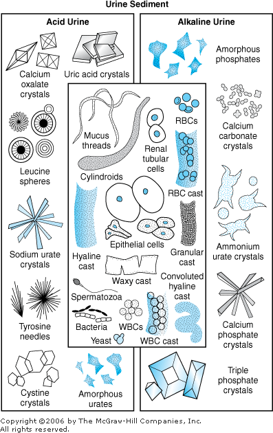 | Urine sediment as seen with a microscope. (Reproduced, with permission, from: Greene MG [ed]: The Harriet Lane Handbook: A Manual for Pediatric House Officers, 12th ed., Year Book Medical Publishers, Chicago, IL, 1991.) |
|
Red Blood Cells (RBCs): Trauma, pyelonephritis, genitourinary TB, cystitis, prostatitis, stones, tumors (malignant and benign), coagulopathy, and any cause of blood on dipstick test (see Differential Diagnosis for Routine Urinalysis, Blood) White Blood Cells (WBCs): Infection anywhere in the urinary tract, TB, renal tumors, acute glomerulonephritis, radiation, interstitial nephritis (analgesic abuse) Epithelial Cells: ATN, necrotizing papillitis. (Most epithelial cells are from an otherwise unremarkable urethra.) Parasites: Trichomonas vaginalis, Schistosoma haematobium infection Yeast: Candida albicans infection (especially in diabetic or immunosuppressed patients or if a vaginal yeast infection is present) Spermatozoa: Normal in men immediately after intercourse or nocturnal emission Crystals  Normal Normal
- Acidic urine: Calcium oxalate (small, square crystals with a central cross; octahedrons), uric acid (rhomboids, hexagons, squares)
- Alkaline urine: Calcium carbonate/phosphate, triple phosphate (struvite, magnesium ammonium phosphate associated with urea-splitting UTI and possible stone formation [coffin lids])
Abnormal - Any: Cystine (colorless hexagons), sulfonamide, leucine (bicycle wheels), tyrosine, cholesterol
- Excessive: Calcium oxylate (excess vitamin C or spinach, ileitis, ethylene glycol poisoning, urolithiasis), uric acid (gout, leukemia, tumor lysis during chemotherapy), triple phosphate (urea splitting UTI and with "infection" stone formation), indinavir (crystals in patients on HIV therapy)
Contaminants: Cotton threads, hair, wood fibers, amorphous substances (all usually unimportant); "dirty urine" may suggest enterovesical fistula Mucus: Large amounts of mucus suggest urethral disease (normal from ileal conduit or continent urinary diversion that uses bowel) or enterovesical fistula. Glitter Cells: WBCs lysed in hypotonic solution Casts: Localizes some or all of the disease process to the kidney itself - Hyaline Casts. Acceptable unless "numerous," benign hypertension, nephrotic syndrome, after exercise
- RBC Casts. Acute glomerulonephritis, lupus nephritis, SBE, Goodpasture disease, aftermath of streptococcal infection (poststreptococcal glomerulonephritis), vasculitis, malignant hypertension
- WBC Casts. Pyelonephritis, acute interstitial nephritis, glomerulonephritis
- Epithelial (Tubular) Casts. Tubular damage, nephrotoxin, virus
- Granular Casts. Breakdown of cellular casts, leads to waxy casts; "dirty brown granular casts" typical for ATN
- Waxy Casts. All cellular casts can become waxy casts. Severe chronic renal disease, amyloidosis
- Fatty Casts. Nephrotic syndrome, DM, damaged renal tubular epithelial cells
- Broad Casts. Chronic renal disease
|
Spot or Random Urine Studies A spot urine, which is often ordered to aid in the diagnosis of various conditions, is done with only a small sample (10 20 mL) of urine. Spot Urine for 2-Microglobulin < 1 mg/24 h or 0 160 g/L (keep sample refrigerated) A marker of renal tubular injury Increased: Diseases of the proximal tubule (ATN, interstitial nephritis, pyelonephritis), viral diseases, drug-induced nephropathy (aminoglycosides), diabetes, trauma, sepsis, HIV, lymphoproliferative and lymphodestructive diseases (multiple myeloma, plasmacytoma) Spot Urine for Cytology Used as an adjunct in the diagnosis of urothelial cancers (primarily transitional cell carcinoma of the bladder, kidney and ureter); limited or no role for renal cell carcinoma. Use a 3-h postvoid and not an AM sample. Spot Urine for Electrolytes Utility is limited because of variations in daily fluid and salt intake; not useful if a diuretic has been taken. - 1. Sodium < 10 mEq/L: Volume depletion, hyponatremic states, prerenal azotemia (eg, CHF, shock), hepatorenal syndrome, glucocorticoid excess
- 2. Sodium > 20 mEq/L: SIADH, ATN (usually > 40 mEq/L), postobstructive diuresis, high salt intake, Addison disease, hypothyroidism, interstitial nephritis
- 3. Chloride < 10 mEq/L: Chloride-sensitive metabolic alkalosis (vomiting, excessive diuretic use), volume depletion
- 4. Potassium < 10 mEq/L: Hypokalemia, potassium depletion, extrarenal loss
Spot Urine for Erythrocyte Morphology The morphology of red cells in a urine sample positive for blood may indicate of the nature of the hematuria. Eumorphic red cells are seen in postrenal, nonglomerular bleeding. Dysmorphic red cells are associated with glomerular causes of bleeding. Labs vary, but > 90% dysmorphic erythrocytes with asymptomatic hematuria indicates a renal glomerular source of bleeding, especially if associated with proteinuria or casts (eg, IgA nephropathy, poststreptococcal glomerular disease, sickle cell disease or trait). If there are 90% eumorphic erythrocytes or even "mixed" results (10 90% eumorphic erythrocytes), a postrenal cause of hematuria requires urologic evaluation (eg, hypercalciuria, urolithiasis, cystitis, trauma, tumors, hemangioma, exercise induced, BPH). Spot Urine for Microalbumin Normal < 30 mcg albumin/mg creatinine (timed collection < 20 mcg/min) Used to determine whether a diabetic patient is at risk of nephropathy or cardiovascular disease. Perform two or three separate determinations over 6 mo to confirm; spot urine preferred. Diabetic patients with a level of 30 300 mcg often need an ACE inhibitor or angiotensin receptor blocker. Six percent of the healthy population has microalbuminuria. The following are the ranges for microalbuminuria recommended by the American Diabetes Association: Spot Urine for Myoglobin Qualitative negative Positive: Skeletal muscle conditions (crush injury, electrical burns, carbon monoxide poisoning, delirium tremens, surgical procedures, malignant hyperthermia), polymyositis Spot Urine for Osmolality 75 300 mOsm/kg, varies with water intake Patients with normal renal function should concentrate > 800 mOsm/kg after 14-h fluid restriction; < 400 mOsm/kg is a sign of renal impairment. Increased: Dehydration, SIADH, adrenal insufficiency, glycosuria, high-protein diet Decreased: Excessive fluid intake, diabetes insipidus, acute renal failure, medications (acetohexamide, glyburide, lithium) Spot Urine for Protein Normal < 10 mg/dL (0.1 g/L) or < 20 mg/dL (0.2 g/L) for a sample taken in the early AM See Differential Diagnosis for Routine Urinalysis, Protein. |
Creatinine Clearance Normal - Men. Total creatinine 1 2 g/24 h; clearance 85 125 mL/min/1.73 m2
- Women. Total creatinine 0.8 1.8 g/24 h; clearance 75 115 mL/min 1.73 m2
- Children. Total creatinine (> 3 y) 12 30 mg/kg/24 h; clearance 70 140 mL/min/1.73 m2 (1.17 2.33 mL/s/1.73 m2)
Decreased: Decreased creatinine clearance results in an increase in serum creatinine usually secondary to renal insufficiency. See Chapter 4, Creatinine, Serum (SCR), for differential diagnosis of increased serum creatinine. Increased: Early DM, PRG Methods for Determination of Creatinine Clearance (CrCl) CrCl is a sensitive indicator of early renal insufficiency. Clearances are ordered for evaluation of patients with suspected renal disease and monitoring of patients taking nephrotoxic medications (eg, gentamicin). CrCl decreases with age; CrCl of 10 20 mL/min indicates severe renal failure and usually the need for dialysis. - 1. Formal 24-h Urinary Collection for Creatinine Clearance. Order a concurrent SCr and a 24-h urine creatinine. A shorter time interval can be used (eg, 12 h), but the formula must be corrected for this change; a 24-h sample is less prone to collection error.
Example: The following are calculations of (a) CrCl from a 24-h urine sample with a volume of 1000 mL, (b) a urine creatinine of 108 mg/100 mL, and (c) a SCr of 1 mg/100 mL (1 mg/dL). 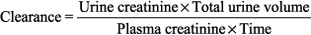

To determine whether there is a valid, full 24-h collection, the sample should contain 18 25 mg/kg/24 h of creatinine for men or 12 20 mg/kg/24 h for women. If the patient is an adult (150 lb = body surface area of 1.73 m2), clearance is not routinely adjusted for body size. Adjustment must be made for pediatric patients. If the values in the previous example are for a 10-year-old boy weighing 70 lb (1.1 m2), the clearance is: 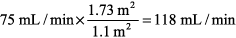
- 2. Estimated Creatinine Clearance. Online calculators for adults and children are available at: www.nkdep.nih.gov/professionals/gfr_calculators (Accessed May 29, 2006)
Adults: - Modification of Diet in Renal Disease (MDRD) equation (Ann Intern Med 1999;130:137 147). The equation does not require weight; results normalized to 1.732 BSA, an accepted adult average BSA.

Cockcroft-Gault equation: 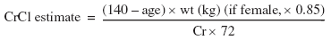
Children: Use the Schwartz equation: 
where k is a constant (0.33, premature infants; 0.45, term infants to 1 y; 0.55, children to 13 y; 0.65, male adolescents; 0.55, female adolescents), height is in centimeters, and SCr is in mg/dL. |
24-Hour Urine Studies Many diseases, most of them endocrine, can be diagnosed with assays of 24-h urine samples. Calcium, Urine Normal: On a calcium-free diet < 150 mg/24 h, average-calcium diet (600 800 mg/24 h) 100 250 mg/24 h Increased: Hyperparathyroidism, hyperthyroidism, hypervitaminosis D, distal RTA (type I), sarcoidosis, immobilization, osteolytic lesions (bony metastasis, multiple myeloma), Paget disease, glucocorticoid excess, immobilization, furosemide Decreased: Medications (thiazide diuretics, estrogens, oral contraceptives), hypothyroidism, renal failure, steatorrhea, rickets, osteomalacia Catecholamines, Fractionated Used to evaluate neuroendocrine tumors, including pheochromocytoma and neuroblastoma. Avoid caffeine and methyldopa (Aldomet) before test. Normal: Values are variable and depend on the assay method used. Norepinephrine 15 80 mg/24 h, epinephrine 0 20 mg/24 h, dopamine 65 400 mg/24 h Increased: Pheochromocytoma, neuroblastoma, epinephrine administration, presence of drugs (methyldopa, tetracyclines cause false increases) Cortisol, Free Used to evaluate adrenal cortical hyperfunction, screening test of choice for Cushing syndrome Normal: 10 110 mg/24 h Increased: Cushing syndrome (adrenal hyperfunction), stress during collection, oral contraceptives, PRG Creatinine See Creatinine, Serum (SCr) and Creatinine Clearance. Cysteine Used to detect cystinuria, homocystinuria, monitor response to therapy Normal: 40 60 mg/g creatinine Increased: Heterozygotes < 300 mg/g creatinine; homozygotes > 250 mg/g creatinine 5-HIAA (5-Hydroxyindoleacetic Acid) 5-HIAA is a serotonin metabolite useful in the diagnosis of carcinoid syndrome. Normal: 2 8 mg /24-h urine collection Increased: Carcinoid tumors (except rectal), certain foods (banana, pineapple, tomato, walnuts, avocado), phenothiazine derivatives Metanephrines Used to detect metabolic products of epinephrine and norepinephrine, primary screening test for pheochromocytoma Normal: < 1.3 mg/24 h for adults, but variable in children Increased: Pheochromocytoma, neuroblastoma (neural crest tumors), false-positive with drugs (phenobarbital, guanethidine, hydrocortisone, MAO inhibitors) Protein See also Urine Protein Electrophoresis. Normal: < 150 mg/24 h Increased: Nephrotic syndrome usually associated with > 3.5 g/1.73 m2/24 h 17-Ketogenic Steroids (17-KGS, Corticosteroids) Overall adrenal function test, largely replaced by serum or urine cortisol levels Normal: Men 5 24 mg/24 h; women 4 15 mg/24 h Increased: Adrenal hyperplasia (Cushing syndrome), adrenogenital syndrome Decreased: Panhypopituitarism, Addison disease, acute steroid withdrawal 17-Ketosteroids, Total (17-KS) Used to measure DHEA, androstenedione (adrenal androgens); largely replaced by assay of individual elements Normal: Men 8 20 mg/24 h; women 6 15 mg/dL. Note: Low values in prepubertal children Increased: Adrenal cortex abnormalities (hyperplasia [Cushing disease], adenoma, carcinoma, adrenogenital syndrome), severe stress, ACTH or pituitary tumor, testicular interstitial tumor and arrhenoblastoma (both produce testosterone) Decreased: Panhypopituitarism, Addison disease, castration in men Vanillylmandelic Acid (VMA) VMA is the urinary product of both epinephrine and norepinephrine; good screening test for pheochromocytoma, also used to diagnose and follow up neuroblastoma and ganglioneuroma Normal: < 7 9 mg/24 h Increased: Pheochromocytoma, other neural crest tumors (ganglioneuroma, neuroblastoma), factitious causes (chocolate, coffee, tea, methyldopa) |
Other Urine Studies Drug Abuse Screen Normal = negative Test for common drugs of abuse, often used for employment screening for critical jobs. Assay varies by facility and may include tests for amphetamines, barbiturates, benzodiazepines, marijuana (cannabinoid metabolites), cocaine metabolites, opiates, phencyclidine. Xylose Tolerance Test (D-Xylose Absorption Test) 5 g xylose in 5-h urine specimen after 25-g oral dose of xylose or 1.2 g after 5-g oral dose Collection: Patient is on NPO status after midnight except for water After 8 AM void, 25 g of D-xylose (or 5 g if GI irritation is a concern) is dissolved in 250 mL water Patient drinks an additional 750 mL water, and urine is collected for the next 5 h. Used to assess proximal bowel function; differentiates malabsorption due to pancreatic insufficiency and that due to intestinal problems Decreased: Celiac disease (nontropical sprue, gluten-sensitive enteropathy), false decrease with renal disease |
Urinary Indices in Renal Failure Use Table 6 1 to differentiate the causes (renal or prerenal) of oliguria. (See also Oliguria and Anuria.) Table 6 1 Urinary Indices Useful in the Differential Diagnosis of Oliguria
|
| | Index | Prerenal | Renal (ATN)a
|
|---|
| Urine osmolality | >500 | <350 | | Urinary sodium | <20 | >40 | | Urine/serum creatinine | >40 | <20 | | Urine/serum osmolarity | >1.2 | <1.2 | Fractional excreted sodiumb
| <1 | >1 | Renal failure index (RFI)c
| <1 | >1 |
|
aAcute tubular necrosis (intrinsic renal failure). 
bFractional excreted sodium: cRenal failure index: 
|
|
Urine Output Although clinical situations vary greatly, the usual, minimal acceptable urine output for an adult is 0.5 1.0 mL/kg/h (daily volume normally 750 2000 mL/d). |
Urine Protein Electrophoresis See Protein Electrophoresis, Serum and Urine and Figure 4 4. |
| | |