167 - Diagnostic Investigation of Mediastinal Masses
Editors: Shields, Thomas W.; LoCicero, Joseph; Ponn, Ronald B.; Rusch, Valerie W.
Title: General Thoracic Surgery, 6th Edition
Copyright 2005 Lippincott Williams & Wilkins
> Back of Book > Appendix
Appendix
Color Plates
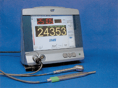 |
Fig. 13-9. Surgical radiation detection system for positrons and 511-keV gamma rays. The console can handle a diverse selection of probes. The high-energy probe shown here has a resolution of 8 mm full width at half maximum (FWHM) at 1 cm. The standard probe commonly use with 99mTc radiotracers has a resolution of 12 mm FWHM at 1 cm. (Node Seeker-800 and PET-Probe. Courtesy of Intra Medical Imaging Inc., Los Angeles, CA, U.S.A.; http://www.gammaprobe.com.) See Fig. 13-9. |
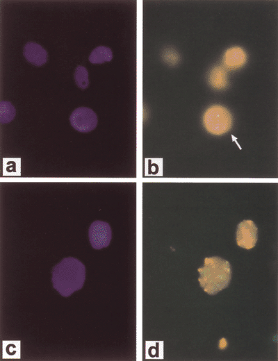 |
Fig. 15-1. Fluorescent in situ hybridization (FISH) demonstrating diploid complement of c-erb-b2/HER-2/neuin normal cells. A. DAPI (4,6-diamidino-2-phenylidole*2HCl) staining of cells. B. FISH (arrow designates diploid signal) and cancer cell line with amplification of c-erb-b2/HER-2/neu. C. DAPI staining of cells. D. FISH. Courtesy of Dr. Paula Capodieci. See Fig. 15-1. |
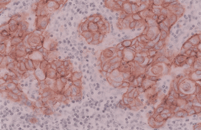 |
Fig. 15-2. Metastatic squamous cell carcinoma to a lymph node with positive staining of HER-2 by immunohistochemistry. See Fig. 15-2. |
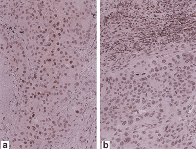 |
Fig. 15-4. Expression of p27 by immunohistochemistry in non small cell lung carcinoma. (A) High expression of p27 in tumor cells. (B) Low expression of p27 in tumor cells with lymphocytes serving as positive internal control. Courtesy of Dr. Michael Murphy. See Fig. 15-4. |
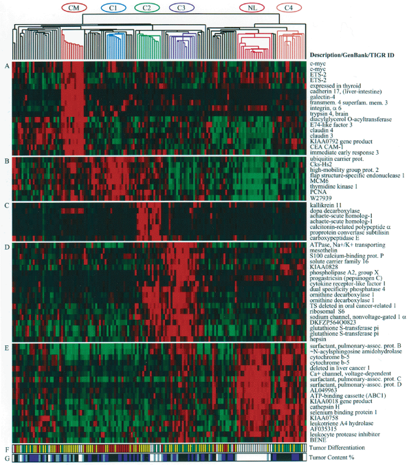 |
Fig. 15-5. Gene expression clusters and histologic differentiation within lung adenocarcinoma subclasses. Genes expressed at high levels in specific subsets of adenocarcinomas. (A) Colon metastases. (B) Proliferation-related gene expression (C1). (C) Neuroendocrine gene expression (C2). (D) Ornithine decarboxylase 1 and surfactant gene expression (C3 and C2). (E) Type II pneumocyte gene expression (C4, C3, and normal lung). (F) Histopathologic degree of differentiation. Red, poor; yellow, moderate; green, well; white, not available or irrelevant. (G) Estimated nucleated tumor content: white, not determined or irrelevant; gray, 30 to 40%; blue, 40 to 70%; black, greater than 70%). Reproduced with kind permission from Bhattacharjee A, et al. Classification of human lung carcinomas by mRNA expression profiling reveals distinct adenocarcinoma subclasses. Proc Natl Acad Sci U S A 98:13790, 2001. See Fig. 15-5. |
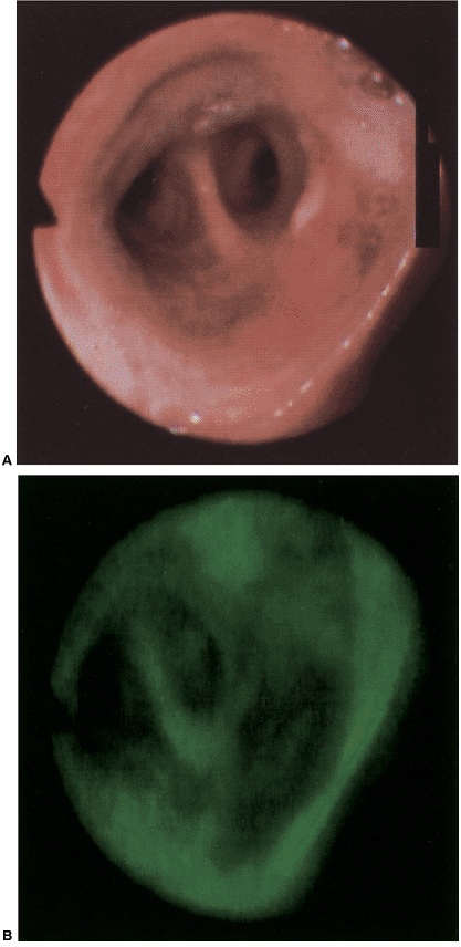 |
Fig. 16-15. A. Photograph of a normal-appearing bronchial mucosa using a white light source. B. Photograph of the same bronchial mucosa using a helium-cadmium laser light source to induce autofluorescence. See Fig. 16-15. |
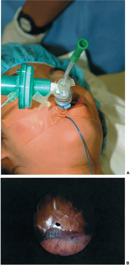 |
Fig. 18-1. Novel technique for selective lobar lung collapse. A. A suction catheter in situ after insertion through a standard endotracheal tube to the target lobar bronchus under guidance of fiberoptic bronchoscopy. B. Needlescopic video-assisted thoracic surgery (VATS) view of selective collapse of the right upper lobe (arrow), with the right lower lobe (foreground) still ventilated. See Fig. 18-1. |
 |
Fig. 18-3. B. VATS exploration confirms the nodule to be sited close to the fissure. See Fig. 18-3. |
 |
Fig. 33-2. The incisions for our approach to video-assisted thoracic surgical lobectomy including the following: (1) an incision for the trocar and the thoracoscope, (2) an incision in the auscultatory triangle for an assistant's instrument, (3) the utility thoracotomy incision, and (4) an incision in the midclavicular line for the stapler. See Fig. 33-2. |
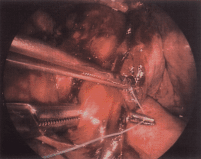 |
Fig. 33-3. A right-angle clamp pulls a tie around the middle lobe bronchus. See Fig. 33-3. |
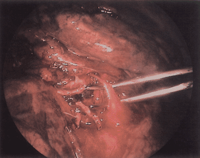 |
Fig. 33-4. A right-angle clamp mobilizes the right middle lobe artery. See Fig. 33-4. |
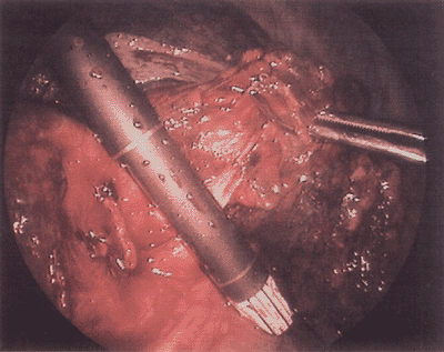 |
Fig. 33-5. The endoscopic stapler is across the right middle lobe artery. See Fig. 33-5. |
 |
Fig. 33-6. The aortopulmonary window lymph nodes are elevated. This exposes the aorta, vagus nerve, and recurrent laryngeal nerve. See Fig. 33-6. |
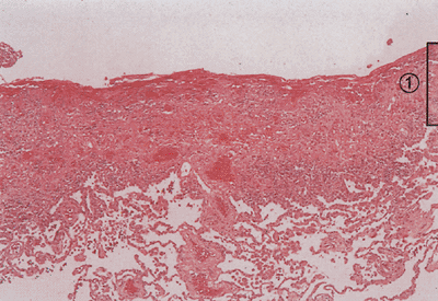 |
Fig. 61-1. Photomicrograph shows that a distinct boundary between early pleuritis (1) and the visceral pleura does not yet exist. Decortication before maturation of the pleural peel will develop a plane between the visceral pleura and pulmonary parenchyma. (Hematoxylin and eosin, original magnification 100.) See Fig. 61-1. |
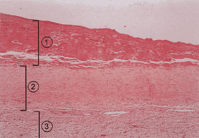 |
Fig. 61-2. Photomicrograph of a mature pleural peel consists of: (1) debris, (2) mature collagen, and (3) a loose vascular matrix overlying the visceral pleura. (Hematoxylin and eosin, original magnification 100.) See Fig. 61-2. |
 |
Fig. 105-2. Regional lymph node stations. See Fig. 105-2. |
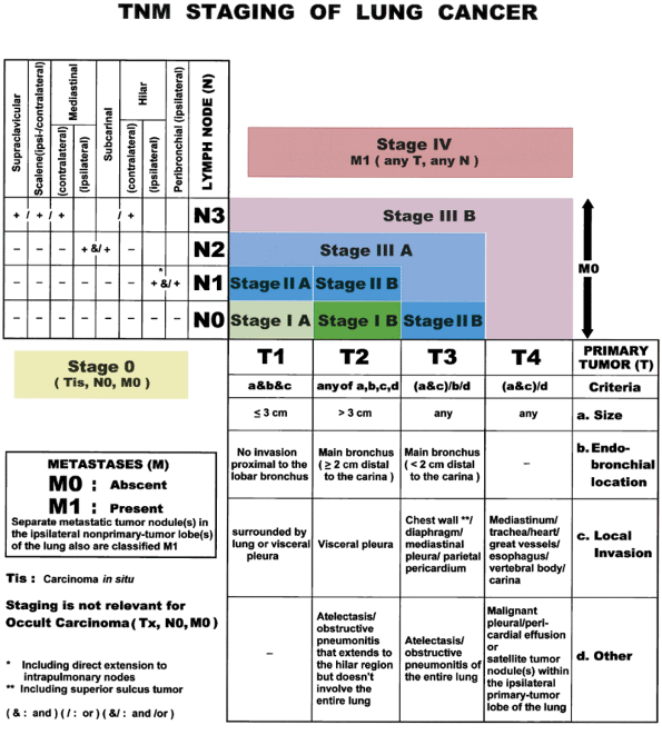 |
Fig. 105-3. TNM staging of lung cancer. From Lababede O, Meziane MA, Rice TW: TNM staging of lung cancer. Chest 115:233, 1999. With permission. See Fig. 105-3. |
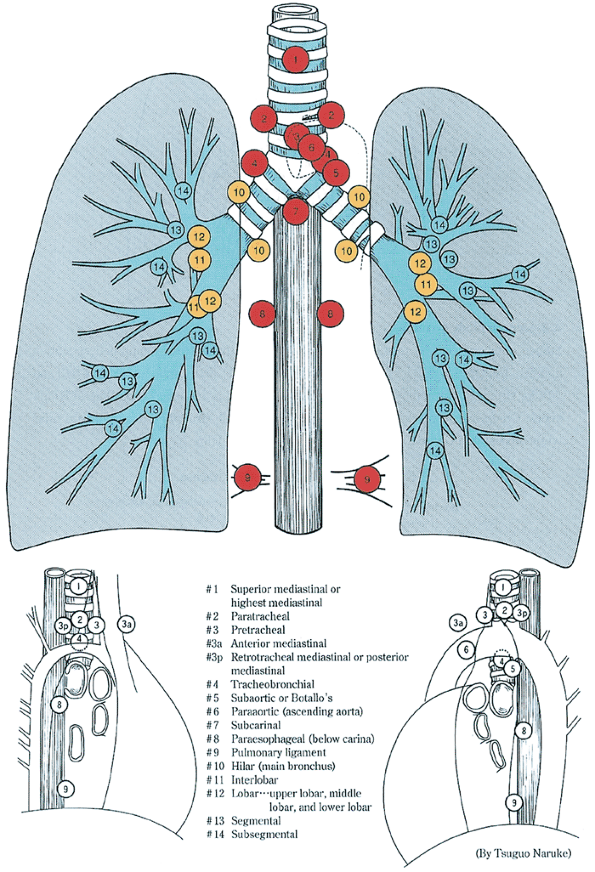 |
Fig. 107-1. A. Lymph node map recommended by the American Joint Committee on Cancer, Union Internationale Contre le Cancer, and the Japan Lung Cancer Society. From Keller SM, et al: Mediastinal lymph node dissection improves survival in patients with stages II and IIIa non-small cell lung cancer. Ann Thorac Surg 70:358, 2000. With permission. See Fig. 107-1. A. |
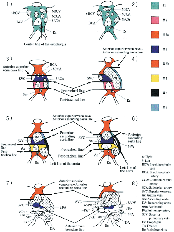 |
Fig. 107-1. B. Sites of mediastinal lymph nodes on computed tomographic scan and cross section. See Fig. 107-1. B. |
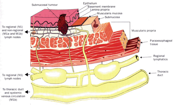 |
Fig. 124-1. Lymphatic anatomy of the esophagus. From Rice T: Superficial esophageal carcinoma: is there a need for three-field lymphadenectomy? Lancet 354:793, 1999. With permission. See Fig. 124-1. |
EAN: 2147483647
Pages: 203