 | Note: Large images and tables on this page may necessitate printing in landscape mode.
Copyright 2007 The McGraw-Hill Companies. All rights reserved.
Clinician's Pocket Reference > Chapter 18. Respiratory Care >
Respiratory Therapy Respiratory therapy is a vital component of health care. For any patient initial medical care begins with assessment of the ABCs: Airway, Breathing, and Circulation. Respiratory therapy includes key components of airway and breathing support. The objective is the care of all types of patients with cardiopulmonary diseases. Functions of the respiratory therapist include emergency care, airway management, ventilatory support, oxygen therapy, humidity and aerosol therapies, chest physiotherapy, physiologic monitoring, and pulmonary diagnostics. |
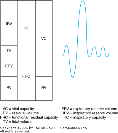
Pulmonary Function Tests (PFTs) PFTs are useful in the diagnosis of a variety of pulmonary disorders. Common PFTs include spirometry, lung volume determinations, and diffusion capacity. Important measurements include FVC and FEV1. Spirometry results indicate the presence of obstructive airway diseases such as asthma and emphysema when the FEV1/FVC ratio is < 0.70. They indicate the presence of restrictive lung diseases such as sarcoidosis and ankylosing spondylitis when the FVC/FEV1 ratio > 80%. Spirometry can be an important part of a preoperative evaluation. Obtain spirograms before and after administration of bronchodilators if they are not contraindicated (ie, history of intolerance). Bronchodilator responsiveness helps in predicting the response to treatment and in identifying asthma. Asthmatic patients typically have at least 15% improvement in FEV1 after bronchodilator therapy. Order lung volumes, commonly determined by helium dilution, to definitively diagnose restrictive lung disease. This test is usually indicated when TLC < 80% of predicted normal value. Diffusion capacity is important in the diagnosis of interstitial lung disease and pulmonary vascular disease, in which it is reduced. Diffusion capacity is frequently monitored to determine response to therapy for interstitial diseases. Obstructive pulmonary diseases include asthma, chronic bronchitis, emphysema, and bronchiectasis. Restrictive pulmonary diseases include interstitial pulmonary disease, diseases of the chest wall, and neuromuscular disorders. Interstitial disease can be caused by inflammatory conditions (usual interstitial pneumonitis [UIP]), inhalation of organic dust (hypersensitivity pneumonitis), inhalation of inorganic dust (asbestosis), and systemic disorders with lung involvement (sarcoidosis). Normal PFT values vary with age, sex, race, and body size. Normal values for a given patient are established from studies of healthy populations and are provided along with the results. ABG should be included in all PFTs. Typical volumes and capacities are illustrated in Figure 18 1. |  | Lung volumes in interpretation of pulmonary function tests. |
|
- Tidal Volume (TV). Volume of air moved during a normal breath on quiet respiration
- Forced Vital Capacity (FVC). Maximum volume of air that can be forcibly expired after full inspiration
- Functional Residual Capacity (FRC). Volume of air in the lungs after a normal tidal expiration (FRC = reserve volume + expiratory reserve volume)
- Total Lung Capacity (TLC). Volume of air in the lungs after maximal inspiration
- Forced Expired Volume in 1 Second (FEV1). Measured after maximum inspiration, the volume of air that can be expelled in 1 s
- Vital Capacity (VC). Maximum volume of air that can be exhaled from the lungs after a maximal inspiration
- Residual Volume (RV). The volume of air remaining in the lungs at the end of a maximal exhalation
|
Differential Diagnosis of PFTs Table 18 1 shows the differential diagnosis of various PFT patterns. When interpreting PFTs, remember that some patients may have combined restrictive and obstructive diseases, such as coexistence of emphysema and asbestosis. | OBSTRUCTIVE AIRWAYS DISEASE (COPD) |
|---|
| Test | Normal | Mild | Moderate | Severe |
|---|
FEV1 (% of VC)
| >75 | 60 75 | 40 60 | <40 | | RV (% of predicted) | 80 120 | 120 150 | 150 175 | >200 | | RESTRICTIVE LUNG DISEASE |
|---|
| Test | Normal | Mild | Moderate | Severe |
|---|
| FVC (% of predicted) | >80 | 60 80 | 50 60 | <50 | FEV1 (% of VC)
| >75 | >75 | >75 | >75 | | RV (% of predicted) | 80 120 | 80 120 | 70 80 | 70 |
|
N = normal;  = increased, = increased,  = decreased; FVC = forced vital capacity; TLC = total lung capacity; RV = residual volume; FEV1 = forced expiratory volume in 1s; VC = vital capacity. = decreased; FVC = forced vital capacity; TLC = total lung capacity; RV = residual volume; FEV1 = forced expiratory volume in 1s; VC = vital capacity. |
|
Oxygen Supplements Table 18 2 describes various methods of oxygen supplementation. Use a bubble-diffuser humidifier to bring the percentage of inspired gas up to room humidity (30 40% of relative humidity [RH]) when using the nasal cannula, simple oxygen mask, partial rebreathing mask, or nonrebreathing mask. Table 18 2 Various Methods of Oxygen and Humidity Supplementation
|
| | Device | O2 Range
| L/min | FIO2
| Uses |
|---|
| Nasal cannula | Low | 1 6 | 0.24 0.5 | COPD, general oxygen needs | | Simple face mask | Medium | 6 8 | 0.5 0.6 | General oxygen needs | | Partial rebreathing face mask | High | 8 12 | 0.6 0.7 | High oxygen emergency needs | | Nonrebreathing face mask | High | 8 12 | 0.7 0.95 | High oxygen emergency needs | | Venturi mask | Low medium | | 0.24 0.50 | COPD (can specify exact FIO2)
|
|
Note: FIO2 vary with fluctuations in the patient's minute ventilation with use of a nasal cannula. This is not true with use of a Venturi mask because it is a "high-flow oxygen enrichment system" that supplies three times the patient's minute ventilation, thus providing an exact FIO2. COPD = chronic obstructive pulmonary disease; FIO2 = fraction of inspired oxygen. |
|
Postoperative Pulmonary Care After a surgical procedure with general anesthesia, the patient needs close observation and good respiratory support (see also Chapter 20). This care includes good suctioning and lateral turning of the head to prevent aspiration. The airway must be patent, and adequate oxygenation and ventilation ensured. For high-risk patients, maintain intubation until it has been clearly determined that the patient can maintain spontaneous ventilation and wakefulness. Determine the capability for maintaining spontaneous ventilation by using the following parameters: - Ability to follow commands
- Vital capacity > 15 mL/kg
- Inspiratory force >~20 cm water
- Expiratory force 25 cm water
- Blood gases with normal PaCO2 (< 44 mm Hg) and pH (> 7.35) during spontaneous breathing
- Adequate PaO2
Administer a judicious regimen of analgesics to control pain but not inhibit the respiratory center. Avoid dressings that restrict chest wall and abdominal excursion. Administer postoperative incentive spirometry, which has been shown effective in decreasing the incidence of postoperative pulmonary complications after upper abdominal operations. Other options include deep breathing exercises and intermittent positive pressure breathing. |
Bronchopulmonary Hygiene The following techniques are used by the respiratory care and nursing services of most hospitals. All are designed to help patients with bronchopulmonary hygiene, more commonly called "pulmonary toilet." Bronchopulmonary hygiene is defined as maintenance of clear airways and removal of secretions from the tracheobronchial tree. This therapy is important for routine postoperative surgical patients, medical patients with obstructive pulmonary diseases, and any patient with excessive respiratory secretions. Aerosol (Nebulizer) Therapy Aerosolized medications such as bronchodilators and mucolytic agents can be delivered via nebulizer to spontaneously breathing, awake patients or intubated patients. Indications - Management of COPD, acute asthma, CF, and bronchiectasis
- Help in inducing sputum for diagnostic tests
Goals - Relief of bronchospasm
- Help in decreasing the viscosity and in clearing of secretions
To Order: Specify the following: - Frequency
- Heated or cool mist
- Medications: In sterile water or NS
- FIO2
- Example: Albuterol 2.5 mg in 3 mL of sterile saline, FIO2 0.28.
Chest Physiotherapy Percussion and postural drainage (P&PD) along with coughing and deep breathing exercises (TC&DB). Position the patient so that the involved lobes of the lung are in a dependent drainage position. Using a cupped hand or vibrator, percuss the chest wall. Nasotracheal (NT) suctioning is quite uncomfortable for patients but is still useful in the appropriate clinical setting in the absence of severe coagulopathy. Indications - Management of pneumonia, atelectasis, and diseases resulting in weak or ineffective coughing
To Order - 1. P&PD: Specify the following:
- Frequency
- Segments or lobes involved (eg, right upper lung [RUL])
- Duration
- Drainage only
- 2. TC&DB: Order on a timed schedule or as needed
- Example: P&PD qid of RUL and RML 5 min/lobe or TC&DB q4h
Incentive Spirometry Encourages the patient to make a maximal and sustained inspiratory effort to help reinflate the lungs or prevent atelectasis. Indications - Treatment of patients at risk of postoperative pulmonary complications
- Management and prevention of atelectasis, especially in the postoperative setting
Goals: Set for the patient depending on the device used: - Lighting lights
- Moving "Ping-Pong" balls
To Order: Specify the following: - Frequency (eg, 10 min q1 2h while patient is awake or 10 repetitions q1h while patient is awake)
- Device (if you have a preference)
- Example: Incentive spirometry 10 repetitions every hour while awake
|
Topical Medications The following agents can be added to aerosol therapy to prevent or manage pulmonary complications caused by bronchospasm, mucosal congestion, and inspissated secretions. Even though these are primarily topical agents, systemic absorption can occur. Acetylcysteine (Mucomyst): A mucolytic agent useful for managing retained mucoid secretions, inspissated secretions, and impacted mucoid plugs that develop in diseases such as COPD, CF, and pneumonia. A bronchodilator should be given along with Mucomyst. Usual Adult Dosage: 1 3 mL of 20% acetylcysteine in 3 mL albuterol Albuterol (Ventolin, Proventil): A short-acting selective bronchodilator with principally beta-2 activity; can cause tachycardia. Onset 15 min after administration. Peak effect at 0.5 1 h, duration 3 5 h. Usual Adult Dosage: 2.5 mg in 3 mL NS q4h Levalbuterol (Xopenex): L-isomer of short-acting albuterol; more expensive but this form causes less tachycardia. Onset 15 min after administration. Peak effect at 0.5 1 h, duration 3 5 h. Usual Adult Dosage: 0.63 1.25 mg in 3 mL NS q6h Racemic Epinephrine: Contains both D- and L- forms of epinephrine. Alpha effects cause mucosal vasoconstriction to reduce mucosal engorgement, and bronchodilation lessens the risk of hypoxemia. Most useful for laryngotracheobronchitis and immediately after extubation in children. Usual Adult Dosage: 0.125 0.5 mL (3 10 mg) in 2.5 mL NS Ipratropium Bromide (Atrovent): A parasympatholytic agent that causes bronchodilation and decreases secretions with "drying" of the respiratory mucosa. Minimally absorbed and rarely causes tachycardia. Onset 45 min after administration. Duration 4 6 h. Usual Adult Dosage: 0.5 mg in 3 mL NS qid Atropine: A parasympatholytic agent; causes bronchodilation and decreases secretions with "drying" of the respiratory mucosa. Readily absorbed and therefore has cardiac effects (tachycardia). Usual Adult Dosage: 0.025 0.05 mg/kg of 1% solution |
Metered-Dose Inhalers (See Chapter 22 for additional prescribing information). All bronchodilating agents can be effectively delivered by metered-dose inhaler as long as the patient can cooperate and proper technique is used. For these devices to be successful, inpatients must be well trained or have the assistance of a nurse or respiratory therapist. Albuterol and ipratropium bromide (Atrovent) can be delivered two puffs q4h. A combination bronchodilator (Combivent) containing the equivalent of one puff of each provides synergistic bronchodilation. |
| |  |