Seeing
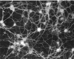
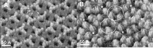
| Perhaps one of the most beneficial tools afforded by the process of realizing the full potential of nanotechnology will be the ability to visualize nanoscalemolecular activity in real time. Continually advancing imaging methods have pushed the boundaries of nanoscale resolution. Fluorescence microscopy greatly enhances the visualization of micro-and nanometer particles in biological specimens. For example, confocal microscopy augments the fluorescent labeling of everything from individual proteins to whole cells by offering several benefits over traditional fluorescent microscopy. Of particular note is that confocal microscopy reduces background fluorescence by using a filter that blocks out fluorescence located outside the focal point of the lens. Because it contains multiple excitation sources, confocal microscopy provides the further benefit of the simultaneous use and visualization of multiple fluorescent dyes. This allows, for example, the visualization of multiple neuronal cell attributes, as illustrated in Figure 17-1. Figure 17-1. Ten-day-old mouse cerebellar neuronal culture labeled with neuronal marker beta III-tubulin and a marker for de novo DNA methyltransferase (Feng, Chang, Li, and Fan 2005). Atomic force microscopy (AFM) has evolved as a valuable method for evaluating surface topography down to the nanoscale, largely because of its incorporation of a flexible cantilever containing a nano-sized tip. Recently, AFM has been used to study nano-sized pores contained in cell membranes that facilitate the exchange of solutes and nutrients between their cytoplasm and their environment. For example, within its outer membrane the bacterium Escherichia coli contains channels called porins that open and close in response to changes in pH and transmembrane voltage. To study the conformational changes of the porins under the influence of pH and voltage, Muller and Engel (1999) employed atomic force microscopy. Upon varying the pH or voltage across the membrane, AFM confirmed conformational changes in the porin, showing a transition from a columnar structure to one consisting of a nano-sized hole, as shown in Figure 17-2. Figure 17-2. High-resolution AFM images of ion gradient and pH-dependent conformation changes of porin OmpF: (A) closed OmpF porin channel; (B) open OmpF porin channel (Müller et al. 1999). Most recently, using materials that can amplify near field optical waves to visualize subjects far below the diffraction limit (Fang et al. 2003; Pendry 2001) will open a new territory for advancing nanotechnologies. The continued enhancement of the nanoscale visualization methods will enable discoveries of complex processes, including receptor-ligand interactions, DNA translocation across cellular membranes, and beyond. Such a capability will revolutionize our ability to directly observe reactions upon which the functionality of future nanotechnological devices will be based. |
EAN: 2147483647
Pages: 204