Angiography
Authors: Flaherty, Alice W.; Rost, Natalia S.
Title: Massachusetts General Hospital Handbook of Neurology, The, 2nd Edition
Copyright 2007 Lippincott Williams & Wilkins
> Table of Contents > Imaging > Angiography
Angiography
A. Diagnostic angiogram
To assess aneurysm, AVM, dural fistula, vessel stenosis, or dissection that is equivocal on CTA/MRA, vessel occlusion before IA thrombolysis, vasculitis, vasospasm, blood flow near a tumor.
B. Interventional angiogram
Aneurysm coiling, intra-arterial thrombolysis, balloon angioplasty, cerebrovascular stenting, mechanical clot retrieval, glue embolization of AVM or tumors, severe epistaxis.
C. Angiographic anatomy
See also Vascular territories, p. 26.
D. Consent
Risk of complication [all equal to] 4%; permanent [all equal to] 1%, death [all equal to] 0.6%. Higher risk with interventional angio, tight stenosis, or h/o migraine.
 |
Figure 18. The circle of Willis. (From Greenberg MS. Handbook of Neurosurgery. 3rd ed. Lakeland, FL: Greenberg Graphics, 1994, with permission.) |
P.176
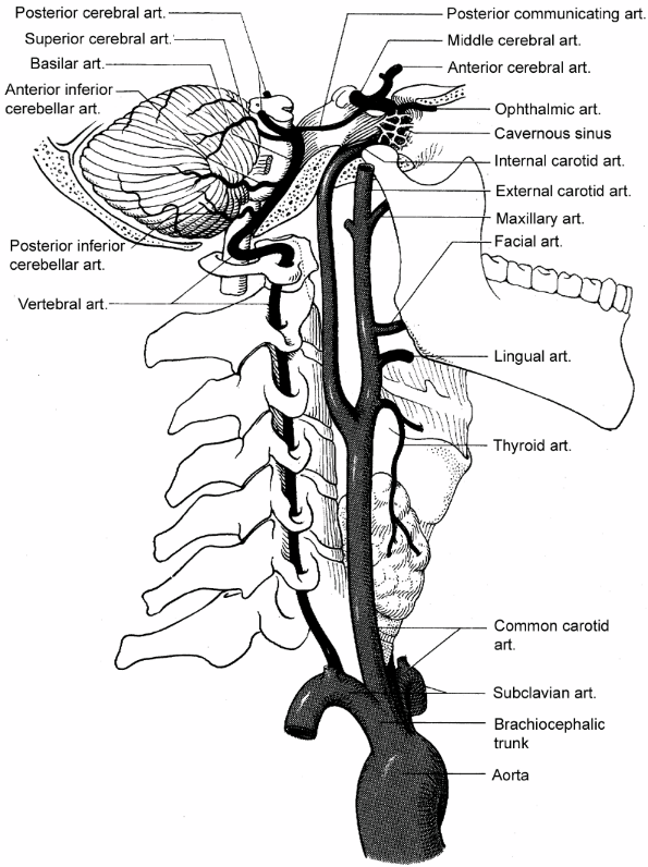 |
Figure 19. Carotid artery anatomy. (Reprinted with permission from Duus P. Topical Diagnosis in Neurology. New York: Thieme, 1983:415.) |
P.177
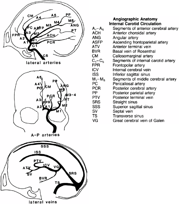 |
Figure 20. The carotid circulation. (From Lerner AJ. The Little Black Book of Neurology. 3rd ed. St. Louis: Mosby, 1995:20, with permission.) |
E. Orders
NPO after midnight, IV fluids, preop labs.
F. Post-angio orders
Keep leg straight for 4 hours; check for groin hematoma and distal leg for pulses (q15min 4, q30min 2, q1h 4). Follow-up noncontrast head CT.
After stenting or mechanical clot retrieval: IIb-IIIa drip, heparin IV, or ASA vs. clopidogrel.
After IA thrombolysis: No anticoagulants or antiplatelets for 24 hrs. Post-tPA precautions, BP control (SBP <180), anticoagulants, or antiplatelets.
After angioplasty or IA treatment of vasospasm etc: IV nicardipine or milrinone.
G. Sheath pull
Usually done at the end of the angiogram; it is sometimes left in longer. Stop anticoagulant drips before pulling sheath. Consider percutaneous closure devices.
P.178
 |
Figure 21. The vertebrobasilar circulation. (From Lerner AJ. The Little Black Book of Neurology. 3rd ed. St. Louis: Mosby, 1995:21, with permission.) |
EAN: 2147483647
Pages: 109