| Note: Large images and tables on this page may necessitate printing in landscape mode.
Copyright 2007 The McGraw-Hill Companies. All rights reserved.
Current Otolaryngology > I. Introduction > Chapter 5. Lasers in Head & Neck Surgery >
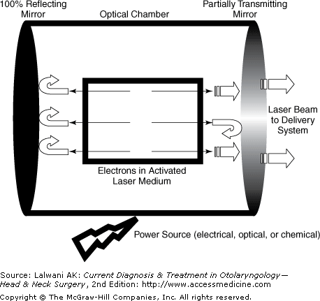
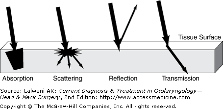
| Definitions The word "laser" is an acronym for L ight A mplification by S timulated E mission of R adiation. A laser is a device that produces an intense beam by amplifying light. Radiation The radiation produced for surgical lasers is in the electromagnetic spectrum with a wavelength that ranges from 200 to 400 nm (near-UV radiation), 400 to 700 nm (visible radiation), 700 to 1000 nm (near-infrared radiation), and more than 1000 nm (infrared radiation). The most prominent physical feature of the radiation is its wavelength, which determines its visibility. The three most commonly used types of surgical lasers are (1) the Argon laser, which is within the visible portion of the electromagnetic spectrum; (2) the neodymium:yttrium-aluminum-garnet (Nd:YAG) laser; and (3) the carbon dioxide (CO 2 ) laser. Amplification Stimulated emission is the main source of laser energy. However, the energy of stimulated emission needs to be amplified to produce an intense beam. When the laser pump activates the active medium, the active medium starts having more atoms in an excited state. As atoms in the excited state release photons, this induces the emission of the photons from other atoms through a chain reaction. Light One of the distinctive features of the light is its highly concentrated energy per unit area. Beams forming the light synchronously occur parallel with each other, which makes it possible for the laser to travel for certain distance without divergence . It is monochromatic . The wavelength of the light is one of the factors determining the physical characteristics of the laser and its interaction with tissue . Stimulated Emission The current model of stimulated emission is described by quantum physics, which defines different energy levels of electrons while revolving around the nucleus in different levels of orbit . In this model, a stable electron in a normal state makes a transition to a higher but unstable energy level by absorbing a photon ( absorption ). This unstable electron with high energy ultimately may return to the original stable level spontaneously ( spontaneous emission). Alternately, this emission can be induced by a forced interaction between one photon and the unstable electron to release a new photon (stimulated emission), which is the basis of laser energy. | | Laser Components A laser primarily consists of three main components: (1) an active medium; (2) a stimulation ( excitation ) mechanism, which is the power source or a laser pump; and (3) an optical chamber (feedback mechanism) (Figure 51). The active medium is the component where the laser radiation is generated. The function of the active medium is to supply a source of stimulated atoms, molecules, and ions. It may be in a solid, gaseous, or liquid state. Different types of lasers are named based on what is used as an active medium. Lasers with a solid state of active medium are the Nd:YAG, ruby, and diode lasers. Lasers using a gaseous active medium are the CO 2 , argon, and helium-neon lasers. The helium-neon laser is used as an aiming beam in lasers with an invisible beam (as in the CO 2 laser) in order to create a visible beam. A laser with a liquid active medium uses organic dye. The activation status of the laser medium is operated by the operation mode of the laser device. Three operational modes are available currently. In the continuous mode, the active medium is kept in a stimulated mode, which provides constant and stable energy. In the pulsed mode, the active medium is intermittently activated for a very short time, which allows tissue to cool off between pulses, thereby decreasing thermal damage. However, a much higher maximum of instantaneous energy is delivered with pulses compared with that of the continuous mode in which average power output is greater. In Q-switched mode, very short pulses of the laser are produced in a controlled manner. The second component of the laser is the power source that is used to activate the medium. The optical chamber is used to direct the output and also to provide feedback from amplification and collimation. The optical chamber contains the active medium. Besides these major components of the laser, it must contain a cooling system, a delivery system, a control unit, and a remote control. Delivery systems are important in the selection of a laser. They can be an articulated arm (for the CO 2 laser), optical fibers (for near-infrared and visible lasers) or a connection between the laser and the operation microscope (for the CO 2 laser). | | Commonly Used Lasers CO 2 Laser The wavelength of the CO 2 laser is 10,600 nm, which is not visible. Its power is between 0.1 and 100.0 W. A cooling system is required to couple to the main system because of the high heat energy produced by the laser. Also, the helium-neon laser beam is used as an aiming beam to make it visible. Its delivery system may be a handpiece at the end of an articulated arm consisting of reflective mirrors, a wave guide, or a micromanipulator to be coupled to an operating microscope. Practically, its energy is absorbed by the tissue within a 0.2-mm depth. Any tissue with a high water content selectively absorbs the CO 2 laser. For an incision, a small spot size with a high power density is preferred. New CO 2 laser systems have a spot size as small as 160  m. For vaporization purpose, a low-power density is applied with a large spot size. This also creates a heat energy that coagulates blood and lymph vessels. However, its hemostasis capability is limited to vessels < 0.5 mm. In skin resurfacing, the CO 2 laser ablates 2060 m of tissue and up to 150 m of residual thermal damage per pass. Generally speaking, the CO 2 laser is used for excision of laryngeal lesions and deep skin resurfacing for rhytids and acne scarring. m. For vaporization purpose, a low-power density is applied with a large spot size. This also creates a heat energy that coagulates blood and lymph vessels. However, its hemostasis capability is limited to vessels < 0.5 mm. In skin resurfacing, the CO 2 laser ablates 2060 m of tissue and up to 150 m of residual thermal damage per pass. Generally speaking, the CO 2 laser is used for excision of laryngeal lesions and deep skin resurfacing for rhytids and acne scarring. Argon Laser The Argon laser is typically used for the coagulation of hemangiomas . Its beam emits a green-blue light, visible in the range of the electromagnetic spectrum (458515 nm) and has a penetration depth of 1 mm. Because of its wavelength, it is almost completely absorbed by hemoglobin, melanin, and myoglobin. Nd:YAG Laser The Nd:YAG laser is a solid-state laser that delivers a 1060-nm beam (near infrared), thereby requiring an aiming beam. The penetration depth is 35 mm because of its low absorption by water and tissue pigments. This low absorption also causes scattering and reflection. Therefore, its use for coagulation purposes requires high power, making the thermal coagulation of vessels and hemangiomas possible. Its delivery system is a fiberoptic carrier, which provides a hemostatic effect at contact. However, it can be used for ablation in a noncontact mode. The Nd:YAG laser is used for tracheobronchial lesions, particularly for its excellent hemostatic qualities; nonablative skin resurfacing; and hair removal in ethnic patient populations. KTP-532 Laser The KTP-532 (potassium titanyl phosphate) laser works by passing an Nd:YAG laser through a KTP crystal, resulting in the emission of half its wavelength (532 nm), which becomes visible. The delivery system is a fiberoptic carrier (for vaporizing and coagulation effects) or a contact quartz tip (for cutting). Since this laser is primarily absorbed by oxyhemoglobin, it is mainly used in the treatment of vascular lesions (including superficial skin lesions and telangiectasias) and the surgical reduction of turbinate tissue. Erbium:YAG Laser The Erbium:YAG laser emits a 2940 W, which is highly absorbed by water (1218 times more efficiently than CO 2 laser). It has the advantage of precise tissue ablation, 520 m per pass, a small zone of residual thermal damage compared with the CO 2 laser. The Erbium:YAG laser has the disadvantage of poor hemostatic qualities and limited collagen tightening compared with the CO 2 laser. It is primarily used for superficial skin resurfacing for fine wrinkles , brown spots, and acne scars. | | Laser-Tissue Interaction The effects of laser on tissue rely on one of the following interactions: absorption, scattering, transmission, or reflection (Figure 52). The type of interaction between a laser beam and any tissue is determined by the wavelength of the laser beam, the operation mode of the laser, the amount of energy applied, and tissue characteristics. The interaction of the laser on tissue can be summarized with the general statement that "the shorter the wavelength, the greater the effect on the tissue." Table 51 shows lasers with their electromagnetic spectrum and penetration depth. Lasers whose wavelengths are within 0.10.8 m (UV and visible region of the spectrum) cause minimal water absorption but considerable hemoglobin-melanin absorption. Lasers with a wavelength > 3 m absorb water. | Table 51. Currently Used Lasers with Their Electromagnetic Spectrum and Penetration Depth.
| | | Electromagnetic Spectrum | Laser Type | Wavelength | Penetration Depth | | Visible lasers | Argon | 514 nm | 0.8 mm | | | KTP-532 | 532 nm | 0.9 mm | | | Flashlamp-Excited Dye | 577 nm | 0.9 mm | | Near infrared lasers | Nd: YAG | 1060 nm | 4.0 mm | | Infrared lasers | Ho: YAG | 2100 nm | 0.4 mm | | | Er: YAG | 2940 nm | 3.0 m | | | CO 2
| 10,600 nm | 30.0 m | |
| The visible lasers penetrate into tissue at approximately 1 mm. However, the Nd:YAG laser goes into tissue at 4 mm and absorbs minimal water. In contrast, the penetration depth of the CO 2 laser is only 30 m, which makes it superb as a cutting tool. Changes in tissue exposed to a laser relate to the temperature created by the laser. In temperatures higher than 50 C, enzymatic activity decreases. Protein denaturation occurs at temperatures over 60 C, the point at which physical changes are seen. However, tissue can still recover with the healing process. Over 80 C, the collagen degrades; 100 C is the temperature at which the water vaporizes, which results in expansion of the steam and finally tissue ablation. Although providing perfect hemostasis, a laser incision causes a delay in wound healing. Collateral thermal damage, though inevitable, can be minimized by using infrared lasers. Lasers can be used for incision, vaporization, or coagulation. Laser beams can be focused to spot sizes < 1 mm in diameter or defocused. A focused beam is used for cutting and a defocused beam for ablation and coagulation. | | Laser Safety Rules Currently, there are two main federal regulations regarding the safe use of the laser in the United States. These are the American National Standard for Safe Use of Lasers (ANSI Z 136.1), which regulates the laser industry, and Safe Use of Lasers in Health Care Facilities (ANSI Z 136.3), which regulates the installation, operation, and maintenance of lasers in health care units. There is one other standard, Safe Use of Lasers in Educational Institutions, which regulates safe use during educational activities (ANSI Z 136.5). Lasers can harm not only the patient but also the surgeon and other personnel in the operating room. Laser system hazards can be related either directly to the effect of the beam on tissue, such as the retina , the corneas, or skin, or secondary conditions such as fire, electrocution, toxic waste, and plume radiation. Beam Hazards A primary concern of unsafe laser use is eye injury . It can occur as a result of direct exposure to either a laser beam or a reflected beam. The best protection is for all personnel to wear approved laser safety glasses . If possible, the patient also needs to wear the same eyewear. If it interferes with the operation field or the procedure, moistened sterile cotton eye pads with moistened towels or metallic eye protectors are needed to cover the eyelids. All operating room windows must be covered with an opaque material at the wavelength of the laser used. An ANSI-approved warning sign at the entrance to all operating rooms should be placed with protective eyewear for those who enter the operating room. Plume Hazards Plume hazards relate to plume radiation and plume content. Plume radiation occurs when the laser beam contacts the smoke plume, which is a by-product of laser beam use. The smoke plume results from heat effect of the laser beam. Some of the energy may shift in wavelength, resulting in secondary emission and often in the visible portion of the spectrum. This secondary emission can cause temporary blindness. Continuous or frequent smoke evacuation is the only solution. Plume content biohazard relates to the direct toxic effect of the plume. In addition to laser smoke elimination , a surgical mask can minimize this risk. Fire Hazards Laser systems may cause airway burns from an endotracheal tube fire when the laser beam strikes an endotracheal tube, which is made of PVC. Therefore, tubes wrapped with reflective tape or made of reflective metals are preferred. Likewise, surgical drapes made of flame-retardant material are advised. The immediate surgical field should be covered with saline-soaked towels. Precautions in Anesthetic Procedures These precautions for anesthetic procedures are important when the operation is performed on the larynx or trachea. A closed ventilatory system that is provided with a small-cuff endotracheal tube is preferred unless the tube obstructs the surgical view. This type of system reduces the possibility of an anesthetic gas leak into the operative field, where the laser beam is present. The cuff may be filled with methylene blue. Ventilation with a high concentration of O 2 and nitrous oxide should be avoided. Jet ventilation is often necessary for glottic and subglottic lesions. American National Standards Institute. For the safe use of lasers. ANSI Z 136.1;2000. (Provides guidance for the safe use of lasers and laser systems.)
| American National Standards Institute. For the safe use of lasers in health care facilities. ANSI Z 136.3;1996. (Provides guidance for the safe use of lasers and laser systems in health care facilities.)
| American National Standards Institute. For the safe use of lasers in educational institutions. ANSI Z 136.5;2000. (Describes control measures for institutions ranging from elementary schools through colleges and universities.)
| Reinisch L. Laser physics and tissue interactions. Otolaryngol Clin North Am. 1996;29:893. (Describes the laser, laser beam, and delivery systems, as well as laser-tissue interactions after briefly reviewing the history of the laser.) [PMID: 8890123]
| Web Sites Laser Institute of America
| www.laserinstitute.org
| (Aims at fostering lasers, laser applications, and laser safety worldwide, offering technical information.)
| | | Lasers in Otology-Neurotology Ear Canal Ear canal lesions that are treatable with lasers include chronic infections, tumors , atresia, scar tissue, and webs. Chronic skin infections of the ear canal may require surgical intervention if medical treatment fails. If the full removal of canal skin is necessary, the argon or KTP-532 laser with low power is used to spot weld the edges or corners of the skin graft , which holds them in position. If whole-skin removal is not required, weeping areas of canal skin can be cauterized under local anesthesia with the argon or KTP-532 laser. Either laser is set at 1.52.0 W with a 1-mm spot size. Polyps or other suspicious soft tissue in the ear canal can be biopsied with the same laser systems. Currently, there is no benefit to using lasers in canal skin carcinoma . Subcutaneous fibrous tissue in acquired ear canal atresia or stenosis can be dissected and vaporized with good hemostasis. This helps to keep the skin of the ear canal intact. In transcanal tympanoplasty, skin incision of the ear canal can be made with the laser without compromising visibility. Tympanic Membrane Fenestration of the tympanic membrane can be performed with a laser (laser-assisted myringotomy) under local anesthesia as an alternative to cold- knife myringotomy under general anesthesia. A CO 2 laser is set at 318 W with a single 100-ms pulse through a hand-held otoscope or 200-mm objective of the microscope. A spot size of 2.02.6 mm is preferred. Diode laser was reported to be effective in this application as well. Myringotomy opening made by either laser may be used for endoscopic examination of the middle ear. Patients with a single attack of otitis media with effusion may be candidates for laser-assisted myringotomy instead of ventilation tube placement. However, short duration (average 15 days) of myringotomy patency and recurrence should be kept in mind for chronic cases. A minimally invasive treatment option was described for tympanic membrane atelectasis and called "laser contraction myringoplasty." In this technique, CO 2 laser set at 0.11 W with a spot size of 0.2 mm is applied to the perimeter of the atelectasis. It has been reported that the laser causes tightening of tympanic membrane tissues, which reduces or eliminates atelectasis. Repairing a tympanic membrane perforation with a laser by spot welding is still far from a routine clinical practice. Nonetheless, in recurrent perforations with unresponsiveness to tympanoplasty, welding the temporalis fascia into the anterior canal skin may be effective. Middle Ear The KTP-532, argon, and CO 2 lasers are useful in middle ear surgery. The CO 2 laser has a small spot size (0.10.2 mm), which provides precision and safe handling. Controversy exists as to which laser system is optimal; each has advantages and disadvantages. Visible lasers ensure the accurate aiming of the laser light. A handpiece eases the manipulation. The light of these lasers can pass through the overlying fluid environment. The spot size and power can vary by changing the distance between the tissue and the probe. The disadvantages of these lasers include the fact that their absorbency depends on pigment, which necessitates cautious work on white bones and tendons because of the possible inner ear damage. Therefore, in revision stapedotomy, visible lasers should not be used. In contrast, there is no inner ear damage risk in using the CO 2 laser given its depth of penetration. However, with the CO 2 laser, the aiming and treatment beams need to be accurately aligned. The lack of penetration through liquid may cause the beam to be weakened if fluid is overlying the target tissue. Visible lasers have better optical precision but less ideal tissue characteristics, and the CO 2 laser has better tissue characteristics but less ideal optical precision. Recently, the Er:YAG laser was found safe in middle ear procedures given its high absorption rate by water, weak penetration through the otic bone, and weak transmission through the perilymph. Laser applications in middle ear surgery are not without complications. Facial nerve injury, severe vertigo, chorda tympani burn, and hearing loss can occur. Small glomus tympanicum tumors can be vaporized with good hemostasis. Visible lasers are advantageous in this application. Although not commonly used, granulation tissue in the mastoid cavity can be removed with the argon laser at 46 W on continuous mode. Laser stapedotomy was first introduced in 1979. Since then, it has gained growing acceptance. Although there are studies showing no difference in the hearing gain between classic and laser stapedotomy, laser stapedotomy appears to be advantageous over classic stapedotomy. Laser stapedectomy eliminates mechanical trauma to the inner ear as well as minimizing the prerequisite fine-hand skills. It provides precision in surgery. Postoperative vertigo is also diminished. Its benefits become more obvious in revision surgery and obliterative otosclerosis. Laser stapedotomy can be performed under local or general anesthesia. The laser is first used on the stapes tendon, and then the posterior crus of the stapes is vaporized. The latter should be done as close to the footplate as possible. Either a rosette pattern of spots with a series of laser shots or 0.6-mm fenestra with a single shot is used in the center of the footplate. After every shot, the char is wiped away. If a series of shots is to be used, the firings of the laser beam should be a few seconds apart. Recommended laser settings are as follows : (1) the spot size is 0.15 mm for the CO 2 laser and 0.20 mm for the KTP-532 and argon lasers; (2) the power is 1.5 W for the CO 2 laser and 1.6 W for the KTP-532 and argon lasers; and (3) the pulse duration is 0.1 s for each. With laser stapedotomy, closure of the air-bone gap within 10 dB is obtained in 9095% of cases. No superiority of any laser system over another is mentioned. For cases with otosclerosis confined to the fissula ante fenestram only, a novel technique has been described, laser stapedotomy minus prosthesis (STAMP). The technique simply includes vaporization of anterior crus first and later anterior one third of the footplate with a hand-held probe of argon laser. Use of a prosthesis is not needed. If the otosclerosis is limited to the fissula ante fenestram only, this should free the remainder of the stapes. If so, the stapedotomy opening is sealed with adipose tissue. The technique may be converted to classic laser stapedotomy in appropriate cases. With this novel technique, it has been stated that high-frequency hearing (68 kHz) was better preserved compared with standard laser stapedotomy and also lasted as long as standard laser stapedotomy. Low incidence of refixation was also noteworthy. In chronic ear surgery, a fixed malleus can be freed from the attic with a laser. Scar tissue, cholesteatomas, and adhesions close to the facial nerve or the stapes can be vaporized and removed. This is one of the most useful applications of the laser in otology. Diseased mucosa or granulation tissue in the mastoid bone can be removed by vaporizing with a defocused laser. A recent study showed that ancillary use of KTP laser significantly improved the rate of cholesteatoma eradication as evidenced in staged intact canal wall surgery. Chronic intractable eustachian tube dysfunction is the new area of interest in laser use. For this condition, endoscopic transnasal laser-assisted surgery (laser eustachian tuboplasty) has been recently described with promising results. In this technique, mucosa and underlying soft tissue of the posterior wall (medial cartilaginous lamina) of the eustachian tube are vaporized through its free border using a 980-nm contact tip diode laser (7 W power, continuous-pulsed mode with 0.2 s on and 0.08 s off) or a wave guide of CO 2 laser (12 W power on superpulse mode and 0.05-s pulse). An interesting area in which the laser is included is laser-Doppler vibrometry, which draws increasing attention in diagnostic otology. The system consists of a helium-neon laser, a joystick-controlled aiming prism both mounted on a microscope, ear speculum-sound coupler assembly with nonreflective glass covering at back, sound generator, and probe-tube microphone. Details of how the system works is beyond the scope of this chapter. The resultant parameter is the umbo velocity, which has been stated to be a useful tool in patients with intact drum to differentiate causes of conductive hearing loss. Inner Ear Until recently, experiences in the inner ear have been limited to the CO 2 and argon lasers, which have been used for the treatment of benign paroxysmal positional vertigo (BPPV). Despite promising results, further clinical experience is necessary to prove the superiority of laser use to the current treatment modalities. A recent animal study was aimed to investigate whether there was any difference in hearing thresholds following cochleostomy that was performed with CO 2 laser, Er:YAG laser, or a microdrill. Comparative results showed that the safest method was microdrill cochleostomy. Some degree of hearing loss, more than with microdrill and less than with Er:YAG laser, resulted from CO 2 laser application. Thus, Er:YAG laser was found to have a greater potential to cause damage. Even classic posterior canal occlusion is reserved for patients unresponsive to repositioning or liberating maneuvers. The occlusion with a laser still does not seem an alternative to these treatment modalities in routine practice. The argon laser is one of the laser systems used in patients with BPPV with the aim of partitioning the posterior semicircular canal. In the procedure, after a mastoidectomy and blue- lining the posterior semicircular canal, the argon laser at 412 W and with a 0.10.5 pulse duration is applied 13 times to create a hole on the canal. The handpiece is held 1 mm away from the canal. After the application, the hole created is covered with the temporalis fascia. It is believed that this application occludes the membranous semicircular canal, which may prevent the cupular movement that results from gravity. The CO 2 laser is also applied directly to the endolymphatic space after removing the bone of the posterior semicircular canal; the opening is then occluded with bone wax. Partial labyrinthectomy and labyrinthine ablation, with the preservation of hearing, have been tried in a very small group of patients with promising results. The argon and CO 2 lasers were used to either weld the open ends of the semicircular canals or ablate the macula. However, this procedure is still not an alternative to labyrinthectomy. Tinnitus is another issue of interest in terms of investigating efficacy of low-power laser treatment. Despite inconsistent results reported, a recent placebo-controlled double-blind study showed that 60-mW laser was not effective in alleviating tinnitus in patients with Mnire disease, presbycusis, and sudden hearing loss. Neurotology Laser systems are not commonly used for tumors in the posterior fossa. The main concern is possible thermal injury of the surrounding structures (the facial nerve and cerebellum) and the transfer of the heat via the cerebrospinal fluid (CSF). However, when taking the necessary measures to protect these structures (ie, covering them with saline-soaked cottonoid), the laser can be used safely for hemostasis on the tumor surface and debulking of the tumor. The KTP-532 (315 W) or CO 2 laser (5 W) with a 1-mm spot size on continuous mode can be an alternative to the ultrasonic surgical aspirator to debulk tumors. Even experienced surgeons prefer accurately applied bipolar coagulation for hemostasis near critical neural structures. Argon and Nd:YAG lasers have some promise for the treatment of vascular tumors. In a vestibular nerve section, the KTP-532 laser at 3 W or CO 2 laser at 1 W is an option to using a scalpel. Anthony PF. Laser applications in inner ear surgery. Otolaryngol Clin North Am. 1996;29:1031. (Reviews previous laser techniques in the inner ear and particularly addresses the use of CO 2 and argon lasers in paroxysmal vertigo and labyrinth surgery.) and argon lasers in paroxysmal vertigo and labyrinth surgery.) [PMID: 8890133]
| Antonelli PJ, Gianoli GJ et al. Early post-laser stapedotomy hearing thresholds. Am J Otol. 1998;19:443. ( Compares hearing results with CO 2 and KTP lasers in patients who have undergone laser stapedotomy.) and KTP lasers in patients who have undergone laser stapedotomy.) [PMID: 9661752]
| Brodsky L, Cook S, Deutsch E et al. Optimizing effectiveness of laser tympanic membrane fenestration in chronic otitis media with effusion: clinical and technical considerations. Int J Pediatr Otorhinolaryngol. 2001;58:59. (Addresses the issues of patient selection, disease factors, and technical factors of lasers.) [PMID: 11249981]
| Buchman CA, Fucci MJ, Roberson JB Jr et al. Comparison of argon and CO 2 laser stapedotomy in primary otosclerosis surgery. Am J Otolaryngol. 2000;21:227. (Compares hearing results obtained with argon and CO 2 laser stapedotomy.) laser stapedotomy.) [PMID: 10937907]
| Hamilton JW. Efficacy of the KTP laser in the treatment of middle ear cholesteatoma. Otol Neurotol. 2005;26:135. ( Presents results of ancillary use of KTP laser in cholesteatoma surgery.) [PMID: 15793394]
| Huber A, Linder T, Fisch U. Is the Er:YAG laser damaging to inner ear function? Otol Neurotol. 2001;22:311. (Investigates effect of Er:YAG laser on air and bone conduction thresholds.) [PMID: 11347632]
| Kakehata S, Futai K, Sasaki A et al. Endoscopic transtympanic tympanoplasty in the treatment of conductive hearing loss: early results. Otol Neurotol. 2006;27:14. (Describes laser-assisted myringotomy and endoscopic transtympanic tympanoplasty through the myringotomy fenestration.) [PMID: 16371841]
| Kiefer J, Tillein J, Ye Q. Application of carbon dioxide and erbium:yttrium-aluminum-garnet lasers in inner ear surgery: an experimental study. Otol Neurotol. 2004;25:400. (Compares hearing thresholds in guinea pigs in which cochleostomy was performed either with CO 2 laser, Er:YAG laser, or microdrill.) laser, Er:YAG laser, or microdrill.) [PMID: 15129125]
| Kujawski OB, Poe DS. Laser eustachian tuboplasty. Otol Neurotol. 2004;25:1. (Describes a novel method to manage chronic eustachian tube dysfunction and presents its results in 56 patients with middle ear atelectasis and effusion.) [PMID: 14724483]
| Nakashima T, Ueda H, Misawa H et al. Transmeatal low-power laser irradiation for tinnitus. Otol Neurotol. 2002;23:296. (Presents no beneficial effect obtained from low-power laser use in tinnitus.) [PMID: 11981384]
| Nissen AJ, Sikand A, Welsh JE, Curto FS. Use of the KTP-532 laser in acoustic neuroma surgery. Laryngoscope. 1997;107:118. (This study reports favorable facial nerve outcome following acoustic neuroma surgery using the KTP-532 laser in a large-case population.) [PMID: 9001275]
| Ostrowski VB, Bojrab DI. Minimally invasive laser contraction myringoplasty for tympanic membrane atelectasis. Otolaryngol Head Neck Surg. 2003;128:711. (Describes a novel technique of CO 2 laser to manage tympanic membrane atelectasis.) laser to manage tympanic membrane atelectasis.) [PMID: 12748566]
| Prokopakis EP, Lachanas VA, Christodoulou PN et al. Implications of laser assisted tympanostomy in adults. Otol Neurotol. 2005;26:361. (Presents outcome of patients with OME and AOM undergoing laser assisted tympanostomy with no ventilation tube placement.) [PMID: 15891634]
| Rhoton AL Jr. Operative techniques and instrumentation for neurosurgery . Neurosurg. 2003;53:907. (Describes laser microsurgery in posterior fossa lesions as well as other special instrumentation.) [PMID: 14519224]
| Rosowski J, Mehta RP, Merchant SN. Diagnostic utility of laser-Doppler vibrometry in conductive hearing loss with normal tympanic membrane. Otol Neurotol. 2003;24:165. (Evaluates efficacy of laser-Doppler vibrometry in differential diagnosis of conductive hearing loss.) [PMID: 12621328]
| Saeed SR, Jackler RK. Lasers in surgery for chronic ear disease. Otolaryngol Clin North Am. 1996;29:245. (Summarizes laser types and reviews their usage in the middle ear for chronic otitis media.) [PMID: 8860923]
| Sedwick JD, Loudon CL, Shelton C. Stapedectomy vs stapedotomy: do you really need a laser? Arch Otolaryngol Head Neck Surg. 1997;123:177. (Comparison of air-bone gap closure and the incidence of sensorineural hearing loss with regard to the use of the drill and KTP and argon lasers to make small fenestra.) [PMID: 9046285]
| Silverstein H, Hoffmann KK, Thompson JH et al. Hearing outcome of laser stapedotomy minus prosthesis (STAMP) versus conventional laser stapedotomy. Otol Neurotol. 2004;25:106. (Compares hearing results in STAMP technique and laser stapedotomy, and concludes better high-frequency hearing preservation in STAMP technique.) [PMID: 15021768]
| Vernick DM. Laser applications in ossicular surgery. Otolaryngol Clin North Am. 1996;29:931. (Reviews laser types and gives their optimal settings in middle ear surgery.) [PMID: 8890125]
| Wiet RJ, Kubek DC, Lemberg P, Byskosh AT. A meta-analysis review of revision stapes surgery with argon laser: effectiveness and safety. Am J Otol. 1997;18:166. (Presents a meta-analysis of hearing results in revision stapes cases which have undergone either non-laser or argon laser surgeries.) [PMID: 9093671]
| Zanetti D, Piccioni M, Nassif N et al. Diode laser myringotomy for chronic otitis media with effusion in adults. Otol Neurotol. 2005;26:12. (Analyzes the closure time of diode laser-assisted myringotomy and recurrence rate of OME.) [PMID: 15699714]
| | | Lasers in Head & Neck Paranasal Sinuses & Nose The introduction of the laser into nasal surgery has resulted from a need for good hemostasis in a narrow operative field. In using the Nd:YAG, KTP-532, and argon lasers, neighboring structures (eg, the medial rectus muscle, anterior cranial fossa, and optic nerve) are at great risk of thermal damage. Therefore, the versatility of the CO 2 and Ho:YAG lasers in this setting have received much recognition. Reducing turbinate hypertrophy and resecting polyps, papillomas, and synechiae can be performed with the CO 2 laser. The physician should be careful not to expose the turbinate bone secondary to thermal damage, which causes scarring, prolonged pain, and persistent crusting. In turbinate resection, the anterior part of the turbinate should always be preserved. The CO 2 laser can be used in superpulse mode to provide gradual vaporization. Using an optical wave guide, the laser beam can be directed at the posterior aspect of the turbinate. During the procedure, the laser plume should be vigilantly evacuated. Although the CO 2 laser provides good hemostasis and less thermal collateral damage in resecting the previously mentioned lesions, the lack of flexibility in its delivery system constitutes a serious problem. The Ho:YAG laser has tissue interaction characteristics similar to the CO 2 laser. It is used on pulse mode. Since it can be transferred through a fiberoptic system, it is more advantageous than the CO 2 laser. Wedge resection of the inferior turbinate using consecutive interstitial and contact beams from an Nd:YAG laser is also efficient. Holmium:YAG laser has been used to correct nasal septal cartilage without elevation of the mucoperichondrial flap. Under local anesthesia using a modified speculum, deviated septum was corrected, and then the laser via an optical fiber was applied through the mucosa. Hereditary hemorrhagic telangiectasia can be undergone with Nd:YAG laser therapy to reduce frequency of epistaxis. It should be noted that, overall, although laser systems offer some advantages, they have not replaced the classic surgical approaches. Katz S, Schmelzer B, Vidts G. Treatment of the obstructive nose by CO 2 laser reduction of the inferior turbinates: technique and results. Am J Rhinol. 2000;14:51. (Presents techniques of the CO 2 laser for inferior turbinate reduction and comparative rhinomanometry results.) laser for inferior turbinate reduction and comparative rhinomanometry results.) [PMID: 10711333]
| Kuhnel TS, Wagner BH, Schurr CP, Strutz J. Clinical strategy in hereditary hemorrhagic telangiectasia. Am J Rhinol. 2005;19:508. (Investigates efficacy of Nd:YAG laser treatment in patients with hereditary hemorrhagic telangiectasia in terms of frequency of nasal bleeding.) [PMID: 16270607]
| Ovchinnikov Y, Sobol E, Svistushkin V et al. Laser septochondrocorrection. Arch Facial Plast Surg. 2002;4:180. (Presents results of nasal septal cartilage reshaping using holmium:YAG laser in 110 patients.) [PMID: 12167077]
| Rathfoot CJ, Duncavage J, Shapshay SM. Laser use in the paranasal sinuses. Otolaryngol Clin North Am. 1996;29:943. (Reviews laser use and its advantages and disadvantages in paranasal sinus surgery.) [PMID: 8890126]
| Vagnetti A, Gobbi A, Algieri GM et al. Wedge turbinectomy: a new combined photocoagulative Nd:YAG laser technique. Laryngoscope. 2000;110:1034. (Describes a new method in inferior turbinate reduction using two consecutive applications of interstitial and contact Nd:YAG laser beams.) [PMID: 10852526]
| Oral Cavity & Oropharynx Laser-Assisted Uvulopalatoplasty The treatment of snoring and sleep apnea is one of the fields in which the laser has gained great popularity. The CO 2 laser has been shown to be an effective treatment instrument for snoring and sleep apnea when the obstruction is at the level of soft palate. Although no difference is found in the postoperative snore index between laser-assisted uvulopalatoplasty (LAUP) and conventional uvulopalatopharyngoplasty, LAUP helps avoid most of the postoperative morbidity, as well as providing a good hemostatic benefit during surgery. However, it has been reported that long-term results of snoring and respiratory disturbance index were not as satisfactory as short- term results and tended to deteriorate over time, which was explained with velopharyngeal narrowing and palatal fibrosis caused by the laser. LAUP can be performed under local-topical or general anesthesia, even in an office setting. The operation can also be staged. The CO 2 laser is the laser most commonly used by otolaryngologists for this operation. Since the diameter of the vessels encountered during the procedure is smaller than 0.5 mm, the CO 2 laser is effective for hemostasis. Basically, in a LAUP, redundant soft tissue is either excised or ablated. In a typical CO 2 laser application, the system is set to a power of 1520 W. A backstop is used to protect the pharynx from scattering the beam. The system is used in the focused mode for excision and in the defocused mode for vaporization. Bilateral incisions at both sides of the base of the uvula are made with a handpiece. The uvula is shortened to 15 mm, excising redundant soft tissue and preserving its curved shape. The wound then heals within 34 weeks. Figure 53 shows a postoperative view of immediately after LAUP. Laser Tonsillotomy Laser tonsillotomy is reserved for patients who are unable to tolerate general anesthesia or unwilling to undergo classic tonsillectomy. The technique requires ablation of tonsillar crypts and gross reduction of tonsillar tissue, which can be staged many times until the level of palatoglossus muscle is achieved. The CO 2 laser is set at 1520 W on continuous mode and applied preferably with a handpiece. Laser Tonsillectomy Laser tonsillectomy is not indicated unless a coagulation disorder is diagnosed because of the cost of the laser. The KTP-532 laser is considered the instrument of choice for tonsillectomy because it provides adequate cutting with good hemostasis and little thermal damage. Its optical fiber is held very close to the tonsillar tissue. The first incision is made in a curvilinear fashion along the anterior pillar from the superior pole to the inferior pole to define the dissection plane. Medially and inferiorly, the retracted tonsil is then dissected from the superior pole to the inferior pole. Lingual Tonsillectomy The excision of the lingual tonsil in the classic fashion is somewhat cumbersome owing to excessive bleeding, postoperative edema, and pain. The use of a laser offers resection with minimal edema, less bleeding, improved visibility during surgery, and less pain postoperatively. The need for a tracheostomy is therefore less likely. The CO 2 , KTP-532, or Nd:YAG laser can be used along with a rigid laryngoscope. The operation can be staged. Benign Lesions CO 2 laser excision or ablation of gingival hyperplasias, pyogenic granulomas, and papillomas is possible with an excellent response rate, good hemostasis, and low morbidity. For especially vascular lesions, photocoagulation with an Nd:YAG laser is preferred. It is set at 3248 W with a pulse duration of 0.3 s. A 2-mm spot size is used with a 2-mm separation between spots. Oral mucositis associated with chemotherapy or radiation therapy may be prevented with low-level laser use. A few mechanisms underlie the healing effect of the laser. The low-level laser has been demonstrated to increase energy production in the mitochondria. It also facilitates conversion of fibroblasts into myofibroblasts from which fibroblast growth factors are released, and these play a role in epithelial repair. The last effect is that of reducing the formation of free oxygen radicals that are stomatotoxic. The most studied form of low-level laser therapy has been helium-neon laser. The CO 2 laser is an alternative. Studies have used low-level laser either prophylactically (before radiation therapy or chemotherapy) or after the appearance of mucosal lesions during the course of radiation therapy or chemotherapy. A recent review showed that even though there is not sufficient evidence to recommend laser use, evidence of its potential usefulness is accumulating . Premalignant Lesions The CO 2 laser is often used for premalignant lesions, including leukoplakia and erythroplakia. Because these lesions are confined to the epithelium, only the superficial layer of the mucosa is removed by leaving 23 mm margins of normal mucosa. The wound is left to granulate and be covered by a new mucosal layer. These lesions can also be ablated. For ablation, a defocused laser at 1020 W with a 100-ms pulse is used. Malignant Lesions The oncologic literature related to head and neck procedures is encouraging with regard to laser use. Compared with traditional methods , the advantages obtained with laser systems are improved visibility, hemostasis, decreased postoperative edema and pain, and better functional results, including speech and swallowing functions. It allows the surgeon to protect the muscular support of the tongue and the floor of mouth. However, no inherent oncologic benefits result from the laser use. Transoral CO 2 laser resection is recommended for superficial T1 and T2 tumors, considering the difficulty in defining the depth of excision with lasers. It is generally accepted that deeply infiltrative tumors, tumors > 4 cm, and tumors involving the maxilla or mandible are not suitable for laser resection. Transoral CO 2 laser excision of oral cavity carcinomas can be performed with the handpiece or a micromanipulator mounted to the operating microscope. Microscopically abnormal tissues can be detected by using 1% toluidine blue. Normal tissue margins generally measure 12 cm beyond the microscopically abnormal tissue. Resection starts by outlining the margins with the CO 2 laser at 6 W and a 100-ms pulse duration. The incision is then made at 10 W on continuous mode. The defect is left for secondary healing or is sutured. The local control rates of T1 and T2 disease addressed with transoral CO 2 laser resection is 80100% at a 2- to 5-year follow-up. The disease-free survival rate is 8388% at a 5-year follow-up. Burkey BB, Garrett G. Use of the laser in the oral cavity. Otolaryngol Clin North Am. 1996;29:949. (Addresses the indications , techniques, results, and complications of laser use in the oral cavity.) [PMID: 8890127]
| Finkelstein Y, Stein G, Ophir D et al. Laser-assisted uvulopalatoplasty for the management of obstructive sleep apnea: myths and facts. Arch Otolaryngol Head Neck Surg. 2002;128:429. (Presents medium- and long-term subjective and objective results of LAUP.) [PMID: 11926920]
| Genot MT, Klastersky J. Low-level laser for prevention and therapy of oral mucositis induced by chemotherapy or radiotherapy. Curr Opin Oncol. 2005;17:236. (Presents literature review on low-level laser use for prevention of oral mucositis.) [PMID: 15818167]
| Kaluskar SK, Kaul GH. Long-term results of KTP/532 laser uvulopalatopharyngoplasty. Rev Laryngol Otol Rhinol. 2000;121:59. (Presents satisfactory results of KTP-laser uvulopalatopharyngoplasty in 84% of the patients who were evaluated in the 4th year after surgery.) [PMID: 10865488]
| Linder A, Markstrom A, Hultcrantz E. Using the carbon dioxide laser for tonsillotomy in children. Int J Pediatr Otorhinolaryngol . 1999;50:31. (Demonstrates the method using the CO 2 laser in tonsillotomy and its results.) laser in tonsillotomy and its results.) [PMID: 10596884]
| Osman EZ, Osborne JE, Hill PD et al. Uvulopalatopharyngoplasty versus laser assisted uvulopalatoplasty for the treatment of snoring: an objective randomised clinical trial. Clin Otolaryngol. 2000;25:305. (Investigates if there was any difference in the snore index following LAUP and UPPP.) [PMID: 10971538]
| Rathfoot CJ, Coleman JA. Laser utilization in the oral pharynx. Otolaryngol Clin North Am. 1996;29:963. (Laser use in the oropharynx is reviewed in a detailed manner from tonsillectomy to malignant disorders.) [PMID: 8890128]
| Saito T, Honda N, Saito H. Advantage and disadvantage of KTP-532 laser tonsillectomy compared with conventional method. Auris Nasus Larynx. 1999;26:447. (Compares pain, intraoperative blood loss, and healing time following conventional and KTP laser tonsillectomy.) [PMID: 10530741]
| White JM, Chaudhry SI, Kudler JJ et al. Nd:YAG and CO 2 laser therapy of oral mucosal lesions. J Clin Laser Med Surg. 1998;16:299. (Describes the use of the contact Nd:YAG and CO 2 lasers in a variety of benign oral lesions.) lasers in a variety of benign oral lesions.) [PMID: 10204434]
| Larynx Laser surgery in the respiratory tract requires additional equipment and safety precautions. Microlaryngeal instruments have a black finish to prevent the reflection or misdirection of the laser beam. A microlaryngoscope with smoke evacuation channels is used. Platforms that act as a backstop have been developed to absorb the laser energy and to prevent the spread of the laser beam down into the trachea. In addition, vocal cord protectors are used to protect the other vocal fold. The delivery systems of various lasers are important considerations in choosing the type of laser for laryngeal surgery. The CO 2 laser can be used through a rigid laryngoscope. The spot size of the CO 2 laser has been reduced to 160 m in new systems when it is used at a distance of 400 mm. This offers better precision and prevents collateral damage. The argon, KTP-532, and Nd:YAG lasers can be transmitted through the laryngoscope to the tissue via a fiberoptic cable. They are not preferred for nonvascular glottic lesions because of excessive energy absorption by the surrounding tissue; however, good results are reported in glottic lesions with the contact Nd:YAG laser. Bilateral Vocal Cord Paralysis The current therapy for bilateral vocal cord paralysis focuses on static airway enlargement procedures at the posterior glottis; these procedures include posterior cordotomy, medial arytenoidectomy, and total arytenoidectomy. The laser provides better hemostasis compared to classic methods. In a posterior cordotomy, laser can be used to incise the vocal cord anterior to the vocal process. The anterior vocal process is then excised or vaporized unilaterally or bilaterally. In a medial arytenoidectomy, the vocal process and medial portion of the arytenoid body are vaporized, preserving the lateral arytenoid body and the aryepiglottic fold. A total arytenoidectomy can also be performed with the CO 2 , KTP-532, and Nd:YAG lasers. Benign Lesions The use of laser for the excision of nodules, polyps, and cysts is not advantageous over microsurgical techniques in terms of preserving uninvolved mucosal layer and lamina propria. However, the surgical precision of the laser has been increased in new systems by adding a microspot manipulator . For these lesions, the laser is set at as low as 4 W of power in the focused mode. As small a spot size as possible should be used. The cautious excision of the lesion with the involved mucosa is necessary. For submucosal lesions, especially cysts and large sessile polyps, a mucosal incision can be made with the laser. The mucosa is then elevated and the lesion is removed in a standard fashion. However, the additional cost of the laser should be taken into account in these cases. For vascular lesions, the laser is far superior to microsurgical interventions in terms of surgical precision and hemostasis. The CO 2 laser is preferred for small vascular lesions such as symptomatic dilated blood vessels and angiomatous clusters of capillaries. During laser surgery for these lesions, and after achieving endoscopic exposure, the defocused laser with a spot size of 300400 m and at 12 W is used with a single pulse of 0.1 s to coagulate the blood supply. This reduces the size of the capillary lesion. The main capillary lesion is then excised with a focused laser at the same level of power. Large vascular lesions are treated with the Nd:YAG laser for palliation. Under endoscopic exposure, a fiberoptic laser cable is introduced and secured. The lesion is then coagulated with the laser in a noncontact mode (a few millimeters away from the lesion) at 20 W and a 0.5-s pulse. The application can be staged in order to observe the response of the lesion and surrounding tissue. Granulation tissue around the arytenoid cartilage, which arises from mucosal defects caused by gastric reflux or sustained mechanical trauma, can be excised with the CO 2 laser when surgery is warranted. The CO 2 laser can also be used to treat superior tracheal granulation tissue. Recurrent respiratory papillomatosis can be ablated with the CO 2 laser, even though it does not provide a better recurrence rate than microlaryngeal surgery. Because the eradication of the virus is not possible, the area of active expression should be addressed. If possible, a 1-mm normal mucosal margin can be included. This intervention should be performed as infrequently as possible to avoid scarring. The laser permits a precise and bloodless excision with less scarring compared with other surgical options. The CO 2 laser can be used for either the excision of bulky disease or superficial vaporization. For excision, a focused laser is set at 4 W with 0.1-s pulse and a 0.5-s pulse interval. The same setting can be used in a defocused mode for superficial vaporization. Recurrences are addressed in the same manner. A comparative study showed that excision by microdebrider was less time-consuming compared with CO 2 laser. Laryngeal stenosis can be addressed with a laser for cutting or coagulating purpose. However, its only advantage over standard treatment methods (ie, scalpel incision and electrocoagulation) is good hemostasis. To prevent reformation , an anterior membranous or thick glottic web can be incised with a CO 2 laser before interposing the tissue flap, keel, or stent placement. In a posterior glottic web, a CO 2 laser is used to incise the arytenoid mucosa (the micro-trapdoor flap) and vaporize the submucosal scar tissue between the arytenoids. Subglottic stenosis < 1 cm in vertical length can also be addressed with a laser to make radial incisions before bronchoscopic dilatation. Malignant Lesions The CO 2 laser offers surgical precision, minimal bleeding, less surgical trauma, and rapid healing in the endoscopic management of carcinoma in situ and early glottic carcinoma. For carcinoma in situ, the local control rate and quality of life obtained in using the CO 2 laser are close to what is achieved with radiation therapy and better than with vocal cord stripping. Also, better ultimate laryngeal preservation is obtained with the CO 2 laser compared with radiation therapy. This holds true when laser CO 2 cordectomy is compared with open surgery. In a typical application, mucosal disease is excised with the CO 2 laser in the superpulse mode at a spot size of 0.50.8 mm. The output power is set to 23 W. If invasion is found in a histopathologic examination, the underlying vocal ligament should also be excised (subligamentous cordectomy), leaving a 1- to 2-mm normal tissue margin. Studies regarding the efficacy of endoscopic laser use for more aggressive disease and tumors invading the anterior commissure are not conclusive. The transoral excision of the supraglottic carcinoma and selected piriform sinus carcinomas can be facilitated with a laser in the context of organ preservation. The neck is addressed in a staged manner. These techniques offer less postoperative morbidity, including the avoidance of tracheotomy and improved swallowing function. Brown DH. The versatile contact Nd:YAG laser in head and neck surgery: an in vivo and clinical analysis. Laryngoscope. 2000;110:854. (Demonstrates comparable tissue and healing effects of the CO 2 and Nd:YAG lasers in rats and presents favorable results obtained with the use of Nd:YAG laser for laryngotracheal lesions and oral-oropharyngeal carcinoma.) and Nd:YAG lasers in rats and presents favorable results obtained with the use of Nd:YAG laser for laryngotracheal lesions and oral-oropharyngeal carcinoma.) [PMID: 10807364]
| Courey MS, Ossoff RH. Laser applications in adult laryngeal surgery. Otolaryngol Clin North Am. 1996;29:973. (Reviews safety measures and laser choice, and its use in vocal cord paralysis and benign and malignant lesions.) [PMID: 8890129]
| Damm M, Sittel C, Streppel M et al. Transoral CO 2 laser for surgical management of glottic carcinoma in situ. Laryngoscope. 2000;10:1215. (Investigates the effectiveness of the CO 2 laser in glottic carcinoma in situ.) laser in glottic carcinoma in situ.) [PMID: 10892699]
| Dedo HH, Yu KC. CO 2 laser treatment in 244 patients with respiratory papillomas. Laryngoscope. 2001;111:1639. (Presents results of frequent excision of respiratory papillomas using the CO 2 laser.) laser.) [PMID: 11568620]
| Eckel HE, Thumfart W, Jungehulsing M et al. Transoral laser surgery for early glottic carcinoma. Eur Arch Otorhinolaryngol. 2000;257:221. (Includes analyses of rates of local control, regional control, organ preservation, and survival in patients with carcinoma in situ and T1 and T2 laryngeal lesions.) [PMID: 10867839]
| El-Bitar MA, Zalzal GH. Powered instrumentation in the treatment of recurrent respiratory papillomatosis: an alternative to the carbon dioxide laser. Arch Otolaryngol Head Neck Surg. 2002;128:425. (Compares the advantages of microdebrider with CO 2 laser in juvenile laryngeal papillomatosis.) laser in juvenile laryngeal papillomatosis.) [PMID: 11926919]
| Gluth MB, Shinners PA, Kasperbauer JL. Subglottic stenosis associated with Wegener's granulomatosis. Laryngoscope. 2003;113:1304. (Evaluates the outcomes of subglottic stenosis in 27 patients with Wegener's granulomatosis with emphasis on CO 2 laser and other treatment modalities.) laser and other treatment modalities.) [PMID: 12897550]
| Maurizi M, Almadori G, Plaudetti G et al. Laser carbon dioxide cordectomy versus open surgery in the treatment of glottic carcinoma: our results. Otolaryngol Head Neck Surg. 2005;132:857. (Analyzes oncologic results in patients with glottic cancers treated by either laser CO 2 or open surgery.) or open surgery.) [PMID: 15944555]
| Rudert HH, Hoft S. Transoral carbon dioxide laser resection of supraglottic carcinoma. Ann Otol Rhinol Laryngol. 1999;108:819. (Describes the use of the CO 2 laser in supraglottic malignant disorders with the intents of cure and palliation, and gives outcome results comparable to the conventional surgery.) laser in supraglottic malignant disorders with the intents of cure and palliation, and gives outcome results comparable to the conventional surgery.) [PMID: 10527270]
| Steiner W, Ambrosch P, Hess CF et al. Organ preservation by transoral laser microsurgery in piriform sinus carcinoma. Otolaryngol Head Neck Surg. 2001;124:58. (Presents the operative technique of transoral excision of piriform sinus carcinoma and its oncologic results with approaches to the neck.) [PMID: 11228455]
| Tracheobronchial System Laser use in the tracheobronchial system is limited to CO 2 and Nd:YAG lasers. In fact, use of the CO 2 laser in bronchoscopy remains restricted by the articulated arm. Another limitation of the CO 2 laser is its hemostatic capability. In contrast, an advantage of the Nd:YAG laser is the ability to use it with both rigid and flexible bronchoscopes. In addition, better hemostasis, even for deeper lesions, is obtained with the Nd:YAG laser. Characteristics of the CO 2 laser confine its use to superficial lesions, including recurrent respiratory papillomatosis involving the tracheotomy site and the trachea, subglottic and tracheal stenosis, and capillary hemangiomas. The use of the CO 2 laser in these lesions is similar to what is described previously for laryngeal applications. For bulky lesions, the Nd:YAG laser is preferred for its vaporization and coagulation effects. In a typical application, the Nd:YAG laser is set at < 30 W and exposure should be kept to < 90 s. Laser use higher than these levels may cause necrosis and perforation in the tracheobronchial wall. Debulking malignant disorders that obstruct the tracheobronchial system can be performed with the CO 2 or Nd:YAG laser for palliation purpose. Photodynamic therapy is useful only for patients with small lesions of squamous cell carcinoma and carcinoma in situ that can be reached with a flexible fiberoptic bronchoscope. Esophagus CO 2 laser has been introduced to endoscopic management of Zenker's diverticulum. The approach has been called "CO 2 laser-assisted diverticulotomy," and it is preferred in rather primary cases. With this approach, a specially designed endoscope with double lips is introduced into the esophageal lumen. At the level of diverticulum, while the anterior lip of the endoscope is directed toward the esophageal lumen, the posterior lip remains at the bottom of the diverticulum, thereby leaving the common wall and cricopharyngeus muscle between the two lips of the endoscope. Then, through an operation microscope with 400-mm lens and CO 2 laser micromanipulator, the common wall is transected with a CO 2 laser set at 510 W on continuous mode. The transection is recommended to continue down to the distal-most part of the common wall. This procedure also transects the hypertonic cricopharyngeus muscle, which is thought to contribute to the pathogenesis of the diverticulum. Thus, both transecting the common wall and relieving the muscle prevent food entrapment. During the procedure, one should be cautious not to violate fascia that envelops the diverticulum. Compared with open technique, this technique reduces operative time. However, one should be aware of the greater possibility of repeated surgery in this approach. Chang CW, Burkey BB, Netterville JL et al. Carbon dioxide laser endoscopic diverticulotomy versus open diverticulectomy for Zenker's diverticulum. Laryngoscope. 2004;114;519. (Describes CO 2 laser use in the management of Zenker's diverticulum and compares the results with those of open technique.) laser use in the management of Zenker's diverticulum and compares the results with those of open technique.) [PMID: 15091228]
| Cholewa D, Waldschmidt J. Laser treatment of hemangiomas of the larynx and trachea. Laser Surg Med. 1998;23:221. (Describes Nd:YAG laser technique and its results in laryngeal and tracheal hemangiomas.) [PMID: 9829433]
| Rebeiz EE, Shapshay SM, Ingrams DR. Laser applications in the tracheobronchial tree. Otolaryngol Clin North Am. 1996;29:987. (Reviews use of the CO 2 , Nd:YAG, and KTP lasers in tracheobronchial tree as well as photodynamic therapy.), Nd:YAG, and KTP lasers in tracheobronchial tree as well as photodynamic therapy.) [PMID: 8890130]
| Savary JF, Monier PF, Fontolliet C et al. Photodynamic therapy for early squamous cell carcinoma of the esophagus, bronchi, and mouth with m-tetra (hydroxyphenyl) chlorin. Arch Otolaryngol Head Neck Surg. 1997;123:162. (Describes the methodology of the photodynamic therapy using m-tetra (hydroxyphenyl) chlorin in carcinoma in situ and microinvasive carcinoma of the upper aerodigestive tract.) [PMID: 9046283]
| Sipila J, Scheinin H, Grenman R. Laser bronchoscopy in palliative treatment of malignant obstructing endobronchial tumors. ORL J Otorhinolaryngol Relat Spec. 1998;60:42. (Describes use of the CO 2 and Nd:YAG lasers with the intent of palliation in malignant endobronchial tumors as well as their fatal complications.) and Nd:YAG lasers with the intent of palliation in malignant endobronchial tumors as well as their fatal complications.) [PMID: 9519381]
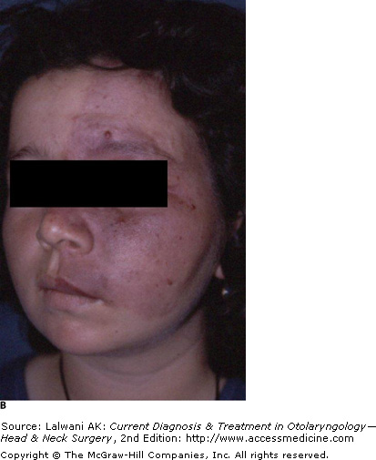
| | | Lasers in Facial Skin Surgery Dermatology is one of the fields in which lasers are most commonly used. Cutaneous lesions present a wide spectrum from vascular lesions to malignant disorders. The use of the laser in dermatology offers surgical precision, improved hemostasis, good preservation of the lesion for histopathologic diagnosis, the facilitation of postoperative wound care, and less scarring. The particular laser selection is based on the histologic nature of the lesion, the lesion site, and the laser characteristics. In dermatology, the cosmetic result is as important as the cure. Patients need to be well informed regarding possible drawbacks of the application, such as a temporary or permanent hypo- or hyperpigmentation, unsightly scarring, and the potential success rate. Skin Resurfacing Ablative Indications for laser surfacing include scars, rhinophyma, actinic cheilitis, superficial squamous cell carcinoma, and wrinkles. For a better result, the depth of thermal damage should be < 100 m. In a typical CO 2 laser application, the pulse duration and power density are adjusted to < 10 ms and 5 J/cm 2 , respectively. With hypertrophic scarring, the scar is ablated with nonoverlapping and intermittent pulses along the lesion. After the application, hyperpigmentation that lasts as long as a few months is expected and is usually reversible. In cases of deep thermal injury, hypopigmentation may occur and is permanent. Because of the significant risk of post-treatment skin dyschromias, CO 2 laser treatment should be limited to lighter- skinned patients. A major advantage of laser resurfacing over classic dermabrasion techniques is less crust formation. Er:YAG laser may be used for superficial resurfacing of fine rhytids and photodamage. Er:YAG laser offers less dramatic change than CO 2 laser with less risk of significant sequelae. Complications reported following laser surfacing are early and late infections by a wide spectrum of agents as well as eruption, prolonged erythema, acne, milia formation, contact dermatitis, hypertrophic scar formation, ectropion, delayed healing, pigmentary abnormalities, inflammatory reactions , and unusual granulomatous reaction. Nonablative Skin Resurfacing Nonablative resurfacing is the use of a laser to induce dermal remodeling without removal of the superficial layers of the epidermis and dermis. Currently, studies have shown minimal improvement in skin quality, tone, and rhytid formation with a variety of Nd:YAG lasers. Rhinophyma In rhinophyma, argon and CO 2 laser systems are alternate options to serial shave incisions and cryosurgery. Compared with serial shave incisions and cryosurgery, under local anesthesia, laser treatments have superior results with better hemostasis. Because the argon laser is absorbed by hemoglobin, the hypervascular form of the disease better responds to the argon laser. The argon laser is set at 1.02.5 W power with a 2-mm spot size and a 0.5-s pulse. A total of 150 pulses is required to treat the entire nose. Treatment sessions should be at least 2 months apart. It takes 10 days to heal after the application. Actinic Cheilitis In actinic cheilitis, CO 2 laser systems with a pulse duration shorter than the thermal relaxation time of the epidermis and dermis provide a better outcome. A typical new CO 2 system for this superficial lesion is set at 250 mJ and 3 W of power at 12 Hz. Conventional CO 2 laser systems with a continuous mode cause thermal damage because they require a longer healing time and may cause more scarring. Vascular Skin Lesions Port-wine stains are the most common vascular lesions. Laser systems are absolutely advantageous in the treatment of these lesions. The goal is to destroy the underlying blood vessels selectively without scarring. For light-colored skin, the flashlamp-excited dye laser is absorbed by red blood cells with minimal absorption in the skin, which causes only minimal thermal damage to epidermis. Its pulse duration is set at 450 s. KTP laser is another alternative. It causes less purpura than flashlamp-excited dye laser. Figure 54A and B show pre- and postoperative views, respectively, of the patient with port-wine stains. The patient underwent three sessions of KTP laser treatment. For dark skin, thrombosis of the vessels is difficult to obtain without damaging the skin because of high melanin absorption. Therefore, infrared lasers are preferred. It should not be used in patients with dark skin or seizure disorders, or in patients receiving anticoagulant or photosensitizing therapy. However, purpura inevitably develops and lasts 1014 days. Temporary or permanent hypopigmentation, transient hyperpigmentation, and scar formation may also develop. In hemangioma and telangiectasia, flashlamp-excited dye is essential to treat the superficial component during both the proliferative phase and the phase of involution of the lesion. Nd:YAG, argon, and KTP-532 laser are other options. Superficial telangiectasias and spider capillaries can be treated effectively with either KTP-532 laser or an intense pulsed light source. Benign Lesions Controversy still exists about whether the CO 2 laser is superior to scalpel excision in treating keloids. However, the advantages of the CO 2 laser to the scalpel include hemostatic superiority and precision when used in the focused mode. In a typical application, a 1-mm spot handpiece is fitted. The laser is set at 10 W in the continuous mode. Excessive tissue is excised with the laser, as with a scalpel. Debris should be cleaned off when necessary; otherwise , the resultant wound would be almost twice as large as the original lesion. The physician should avoid using sutures. However, until reepithelization occurs, the wound should be watched closely. Caf au lait maculas and lentigines are the most common benign lesions. With these lesions, cosmetically better outcomes are obtained with laser systems compared with scalpel excision. Shorter wavelength lasers are preferred because of the pigment content of the lesions. Q-switched laser systems (eg, the pulsed dye laser of 504 nm, or the ruby or Nd:YAG lasers) are ideal for targeting pigmented cells. The CO 2 laser is another option in spite of its much longer wavelength. In terms of scar formation and healing time, CO 2 laser systems with a short pulse duration (200-ms pulses at 250 Hz and 80-W power) provide slightly better outcomes compared with conventional continuous CO 2 systems. Facial verrucae and rosacea are also successfully treated with flashlamp-excited dye laser. Malignant Lesions Basal cell carcinoma, squamous cell carcinoma, and melanoma are the three most common malignant lesions encountered. Laser use is one option among scalpel excision, Mohs micrographic excision, and radiation therapy. The CO 2 laser is ideal, especially for small to moderate lesions. It is also advantageous for use in patients with coagulation defects. In addition, the CO 2 laser is good in preserving margins. The recommended margin of excision is 47 mm for basal cell carcinoma, 34 mm for squamous cell carcinoma, and 13 cm for melanoma. | | Laser-Assisted Hair Removal Lasers in hair removal induce selective damage to hair follicles, while avoiding the competing chromophobe of melanin. Temporary hair reduction is a delay in hair growth, typically lasting 13 months. Permanent hair reduction reduces the number of terminal hairs after a given treatment, usually lasting 6 months. Complete hair loss is the reduction of number of regrowing hairs to zero. Lasers initially produce complete but temporary hair loss. Eventually, the laser creates partial but permanent hair loss (a permanent reduction in the total number of terminal hairs). In patients with light skin, the 694-nm ruby laser and 755-nm Alexandrite laser are used. In patients with darker skin, 800-nm diode laser, 1064-nm Nd:YAG laser (long-pulse and Q-switched), and intense pulsed lights are favored because of less competition with melanin. Alora MB, Anderson RR. Recent developments in cutaneous lasers. Lasers Surg Med. 2000;26:108. (Describes developments in skin cooling between laser applications, laser use in vascular and pigmented lesions, laser resurfacing, and hair removal.) [PMID: 10685084]
| Alster TS, Lupton JR. Prevention and treatment of side effects and complications of cutaneous laser resurfacing. Plast Reconstr Surg. 2002;109;308. (Describes potential side effects of laser resurfacing and management.) [PMID: 11786830]
| Dijkema SJ, van der Lei B. Long-term results of upper lips treated for rhytides with carbon dioxide laser. Plast Recontr Surg. 2005;115:1731. (Presents an evaluation of long-term outcome of CO 2 laser use in upper lips for rhytides.) laser use in upper lips for rhytides.) [PMID: 15861082]
| Doctoroff A, Oberlender SA, Purcell SM. Full-face carbon dioxide laser resurfacing in the management of a patient with the nevoid basal cell carcinoma syndrome. Dermatol Surg. 2003;29:1236. (Presents CO 2 laser use in a particular case with multiple basal cell carcinoma on the face.) laser use in a particular case with multiple basal cell carcinoma on the face.) [PMID: 14725671]
| Lonne-Rahm S, Nordlind K, Edstrom DW et al. Laser treatment of rosacea. Arch Dermatol. 2004;140:1345. (Presents results of flashlamp pulsed dye laser use in 32 patients with rosacea.) [PMID: 15545543]
| Rendon-Pellerano MI, Lentini J, Eaglstein WE et al. Laser resurfacing: usual and unusual complications. Dermatol Surg. 1999;25:360. (Presents early and late complications in seven cases of patients who have undergone laser skin resurfacing.) [PMID: 10469072]
| Ries WR, Speyer MT. Cutaneous applications of lasers. Otolaryngol Clin North Am. 1996;29:915. (Describes particular laser selection and laser use in a wide spectrum of dermatologic lesions from keloid and pigmented lesions to malignant lesions.) [PMID: 8890124]
| Vargas H, Hove CR, Dupree ML et al. The treatment of facial verrucae with the pulsed dye laser. Laryngoscope. 2002;112:1573. (Describes pulsed dye laser use in facial verrucae.) [PMID: 12352665]
| Waner M. Recent developments in lasers and the treatment of birthmarks. Arch Dis Child. 2003;88:372. (Presents advances in management of port wine stains, hemangiomas, and other pigmented lesions.) [PMID: 12716698]
| | | | |
 Print Close Window