1 - Acute Resuscitation

Editors: Susla, Gregory M.; Suffredini, Anthony F.; McAreavey, Dorothea; Solomon, Michael A.; Hoffman, William D.; Nyquist, Paul; Ognibene, Frederick P.; Shelhamer, James H.; Masur, Henry

Title: Handbook of Critical Care Drug Therapy, 3rd Edition

Copyright 2006 Lippincott Williams & Wilkins

> Table of Contents > Chapter 1 - Acute Resuscitation

Chapter 1

Acute Resuscitation

TABLE 1.1. Advanced Cardiac Life Support (ACLS) Drugs

Drug Dosage Indications/Comments
Antiarrhythmic Agents
Adenosine Bolus: 6 mg (initial)
If no response, bolus with 12 mg after 1 2 min
For conversion of PSVT unresponsive to vagal maneuvers
  May repeat 12 mg once Give as rapid IV bolus over 1 3 s followed by rapid 10 ml fluid bolus
May use lower bolus dose of 3 mg if central line available
Be cautious of interactions with theophylline (inhibits adenosine), dipyridamole (potentiates adenosine), other drugs that prolong QT interval
Amiodarone 300 mg bolus IV
150 mg bolus IV followed by 1 mg/min for 6 h and then 0.5 mg/min for 18 h
For VF or pulseless VT refractory to shock; may repeat 150 mg bolus IV in 3 5 min
For stable wide-complex tachycardia up to a total dose 2.2 g IV per 24 h
May use for narrow complex atrial arrhythmias, as adjunct to cardioversion
Monitor for bradycardia and hypotension
Atropine Bolus: 0.5 mg IV (maximum dose 3 mg) For either absolute (<60 beats/min) or relative symptomatic bradycardia
  Bolus: 1 mg IV For bradycardia manifesting with lack of pulse, PEA or for asystole unresponsive to epinephrine
May repeat dose every 3 5 min up to maximum dose 0.04 mg/kg or 3 mg
  ETT bolus: 2 3 mg Dilute up to 10 ml in NS or sterile water
(IV preferred)
Diltiazem 0.25 mg/kg IV bolus over 2 min (typically 15 20 mg) For control of ventricular response rate in A fib or A flutter, or other narrow complex tachycardia
  May repeat once Do not use in wide-complex tachycardia
  0.35 mg/kg IV bolus over 2 min (typically 25 mg) Negative inotrope, so use cautiously if reduced LV function
  Maintenance infusion 5 15 mg/h  
Epinephrine Bolus: 1 mg IV (10 ml of 1:10,000 solution) Therapy for refractory VF or pulseless VT; dose should be followed by CPR and defibrillation; may be repeated every 3 5 min
    Initial therapy for PEA; may repeat every 3 5 min
    Initial therapy for asystole; may repeat every 3 5 min
  ETT bolus: 2 2.5 mg Dilute up to 10 ml NS or sterile water
(IV preferred)
  Infusion: 2 10 g/min For treatment of symptomatic bradycardia unresponsive to atropine and transcutaneous pacing; alternative to dopamine
Ibutilide 1 mg IV infused over 10 min; may be repeated after 10 min
Use 0.01 mg/kg if <60 kg
For treatment of atrial arrhythmias
Monitor electrolytes and EKG
Increased risk for torsade de pointes if elderly, abnormal LV function (EF <35%), or electrolyte abnormalities
Monitor for 4 24 h
Isoproterenol Infusion: 2 10 g/min May be used in torsade de pointes unresponsive to magnesium
    Use with extreme caution; at higher doses is considered harmful
    Not indicated for cardiac arrest, hypotension, or bradycardia
Lidocaine Bolus: 1 1.5 mg/kg For wide-complex tachycardia of uncertain type, stable VT, and control of PVCs
May be followed by boluses of 0.5 0.75 mg/kg every 5 10 min up to a total of 3 mg/kg
Only bolus therapy should be used in cardiac arrest
  Bolus: 1.5 mg/kg Initial bolus dose suggested when VF is present and defibrillation and epinephrine have failed
  ETT bolus: 2 4 mg/kg Diluted in 5 10 ml NS or sterile water
(IV preferred)
  Infusion: 2 4 mg/min Continuous infusion used after bolus dosing and following return of perfusion to prevent recurrent ventricular arrhythmias
Because half-life of lidocaine increases after 24 48 h, the dose should be reduced after 24 h, or levels should be monitored
Therapeutic levels 1 4 mg/L
Full-loading dose but reduced infusion rate in patients with low cardiac output, hepatic dysfunction, or age over 70 years
Magnesium sulfate Bolus: 1 2 g (8 16 mEq) Drug of choice in patients with torsade de pointes
For recurrent/refractory VT or VF
For hypomagnesemia
For ventricular dysrhythmias, administer over 1 2 min
For magnesium deficiency, administer over 60 min
  Infusion: 0.5 1 g/h (4 8 mEq/h) Rate and duration of infusion determine clinically or by magnitude of magnesium deficiency
Naloxone 0.4 mg IV is typical Onset of action 2 min IV and <5 min IM/SC
  May give 0.4 2 mg IV every 2 3 min (maximum dose is 10 mg) Duration of action ~45 min
Give 0.4 mg diluted in 10 ml NS or sterile water slowly to avoid abrupt narcotic withdrawal
  0.8 mg IM/SC Hypertension/hypotension, cardiac arrhythmias, pulmonary edema may occur
Monitor for reoccurring respiratory depression because narcotics typically last longer than naloxone
  ETT: 2 mg diluted in 5 10 ml NS or sterile water (IV preferred)
Procainamide 12 17 mg/kg; administer at rate of 20 30 mg/min (maximum 50 mg/min) Infrequently used
Recommended when lidocaine is contraindicated or has failed to suppress ventricular ectopy
Use higher dose for more urgent situations (VF or pulseless VT)
Maximum total dose of 17 mg/kg
Continue bolus dosing until arrhythmia suppressed, hypotension, QRS complex widens by 50% of original width, or maximum total dose given
Rapid infusion may cause precipitous hypotension
Avoid in patients with QT prolongation (>30% above baseline) or torsade de pointes
  Infusion: 1 4 mg/min Continuous maintenance infusion, after return of perfusion, to prevent recurrent arrhythmias
Reduce dosage in renal failure
Monitor blood levels in patients with renal failure or with >24-h infusion
Therapeutic levels: procainamide 4 10 mg/L, N-acetyl-procainamide (NAPA) 10 20 mg/L
Vasopressin 40 U IV push, one dose only As an alternative to 1st or 2nd dose epinephrine in refractory VF, asystole, or PEA resume epinephrine after 3 5 min
Verapamil Bolus: 2.5 10 mg over 2 3 min Only give to patients with narrow complex PSVT unresponsive to adenosine
  May repeat in 15 30 min prn
Max. cumulative = 20 mg
Diltiazem (0.25 mg/kg) is an alternative to verapamil because it has less negative inotropy
Vasopressor Agents
Dopamine (For other vasopressors, Table 3.8) Infusion: 2 20 g/kg/min For treatment of symptomatic bradycardia unresponsive to atropine and transcutaneous pacing
For treatment of hypotension that is unresponsive to volume
Electrolyte Agents
Sodium bicarbonate Bolus: 1 mEq/kg Helpful in limited clinical conditions: hyperkalemia, bicarbonate responsive acidosis, tricyclic antidepressant overdose
Not recommended in the majority of arrest cases (hypoxic lactic acidosis)
Guide therapy by blood gas analyses and calculated base deficit to minimize iatrogenic alkalosis
A fib, atrial fibrillation; A flutter, atrial flutter; CPR, cardiopulmonary resuscitation; EF, ejection fraction; EKG, electrocardiogram; ETT, endotracheal tube; IM, intramuscular; IV, intravenous; LV, left ventricular; MI, myocardial infarction; NS, normal saline; PEA, pulseless electrical activity; PSVT, paroxysmal supraventricular tachycardia; PVC, premature ventricular contraction; SC, subcutaneous; VF, ventricular fibrillation; VT, ventricular tachycardia

P.5


P.6


TABLE 1.2. Shock General Management

Type of Shock Initial Therapy Subsequent Therapy
Cardiogenic Shock
Massive myocardial infarction Supplemental oxygen, aspirin, pain relief, venous access
Therapy for ACS (see Table 3.1)
Optimize volume status and ensure adequate preload
Treat arrhythmias
Consider RHC
Determine need for inotropic agents; diuretics; vasodilators; vasopressors (see Tables 3.3 and 3.8)
Consider mechanical ventilation
Early IAB, coronary arteriography, and revascularization by PCI or bypass grafting
Consider thrombolytic agent if cardiac catheterization not possible
Nonischemic cardiomyopathy Therapy as above, but omit therapy for ACS Consider IAB or assist devices as bridge to transplantation
Consider reversible causes (e.g., acute valvular regurgitation requiring emergent valve replacement, thyrotoxicosis)
Oligemic Shock
Massive hemorrhage, severe dehydration, etc. For hemorrhage, large bore peripheral or central venous access
Volume resuscitation with packed RBCs and 0.9% NaCl
Consider use of blood warmer
If large bleed, consider platelets, fresh frozen plasma, and supplemental calcium
For dehydration, volume resuscitation with 0.9% NaCl or Ringer's lactate
Monitor electrolytes and coagulation
If hypotension persists despite volume resuscitation, consider: possibility of coexisting sepsis, tamponade, or ACS; RHC; inotropic and/or vasopressor agents
Consult GI and surgery for massive gastrointestinal hemorrhage (see Table 7.1)
Extracardiac Obstructive Shock
Tamponade Confirm suspected diagnosis with echocardiography and/or RHC, temporize by increasing filling pressures with bolus 0.9% NaCl IV; support BP Urgent percutaneous pericardiocentesis, surgical pericardiotomy, or pericardial window
Massive pulmonary embolism Correct hypoxemia; administer heparin or LMWH; thrombolytic therapy (alteplase 100 mg IV over 2 h); give inotropic support such as dobutamine for right heart strain and failure Consider percutaneous catheter suction thrombectomy or thoracotomy with embolectomy
Consider IVC filter long term
See Table 4.11
Tension pneumothorax Emergent needle or tube thoracostomy Tube thoracostomy
Distributive Shock
Septic shock Emergent broad-spectrum antibiotics IV after blood cultures; IV crystalloid; if shock persists, consider RHC
If shock persists despite adequate preload, add dopamine 2 20 g/kg/min; or norepinephrine 2 g/min
Consider vasopressin in refractory shock
Stress dose steroids hydrocortisone 100 mg IV q8h; optional fludrocortisone 50 g po qd
Consider baseline cortisol level prior to glucocorticosteroid therapy and corticotropin stimulation test
Role of drotrecogin alfa is not establisheda
Anaphylaxis Epinephrine 0.3 0.5 mg for severe symptoms of hypotension, bronchospasm, or laryngeal edema; given as 0.3 0.5 ml of 1:1,000 SC or 0.5 1.0 ml of 1:10,000 solution IV; also give diphenhydramine 50 mg IV; repeat 25 50 mg IV q4h prn; abnormal permeability causes intravascular depletion, which should be corrected with volume Cautious administration prn of additional epinephrine; give corticosteroids (Methylprednisolone 60 mg IV or equivalent) and cimetidine 300 mg IV q12; these will have delayed rather than immediate effect
For persistent symptoms and patient on -blockers, give glucagon 1 mg IV
Hypoadren-alism Administer dexamethasone 4 mg IV q6h together with fluids To confirm diagnosis, perform corticotropin stimulation test (dexamethasone will not interfere); draw baseline cortisol level, give 250 g IV cosyntropin, and repeat cortisol level 30 min later
Neurogenic Trendelenburg position; fluids If hypotension persists, consider vasopressors (e.g., phenylephrine or metaraminol)
ACS, acute coronary syndrome; BP, blood pressure; GI, gastroenterology; IAB, intra-aortic balloon; IV, intravenous; IVC, inferior vena cava; LMWH, low molecular weight heparin; PCI, percutaneous coronary intervention; RBCs, red blood cells; RHC, right heart catheterization
aWhile some authors recommend drotrecogin alfa (recombinant activated protein C) in highly selected patients with a high risk of death (Apache score of 25) and a low risk of bleeding, the role of drotrecogin alfa in septic patients has not been clearly established. It has no effect or is harmful in septic patients with Apache score of <25, in surgical patients with single organ dysfunction, and in pediatric sepsis. The risk of serious bleeding including intracerebral hemorrhage is increased in patients receiving drotrecogin alfa.

P.7


TABLE 1.3. Hypovolemic Shock

  Mild Moderate Severe Life-threatening
% loss of intravascular volume 10 15% 15 30% 30 40% >40%
Loss of intravascular volume (cc) <700 800 800 1500 1500 2000 >2000
Mean arterial pressure WNL WNL Reduced Reduced
Heart rate 80 100 101 119 120 140 >140
Pulse pressure WNL/increased 101 119
Reduced
120 140
Reduced
>140
Reduced
Respirations (breaths/min) 15 20 21 29 30 35 >35
Capillary refill testa 2 s >3 s >3 s >3 s
Urine output (cc/h) 30 20 30 5 15 Oliguria
Mental status Uneasy Mild anxiety Anxiety or confusion Confusion or lethargy
Volume replacement Crystalloid Crystalloid Crystalloid or blood if indicated Crystalloid or blood if indicated
aThe capillary refill test is performed by pressing on the fingernail or the hypothenar eminence. The test is not valid in hypothermic patients.
WNL = within normal limits.

P.8


TABLE 1.4. Crystalloids and Colloids

Fluid Dosage Comments
0.9% NaCl 500 mla Hyperchloremic metabolic acidosis secondary to vigorous NaCl replacement may occur
Lactated Ringer's 500 mla Balanced electrolyte composition (mEq/L): Na+ 130, K+ 4, Ca++ 3, Cl- 110, lactate 28
Not compatible with blood products
5% albumin 0.25 1 g/kgb Each 250 ml contains 12.5 g albumin
25% albumin 0.25 1 g/kgb Each 100 ml contains 25 g albumin
6% hetastarch 500 mla Chemically modified glucose polymer
Large doses, especially >1500 ml, can lead to coagulopathies (factor VIII deficiency) and platelet abnormalities
Can cause artifactual hyperamylasemia
Anaphylactoid reactions have occurred
Total amount should not exceed 1,500 ml/d (20 ml/kg)
Cautious use in cardiac bypass and septic patients
6% hetastarch in lactated electrolyte injection 500 1,000 ml Chemically modified glucose polymer
Balanced electrolyte composition (mEq/L): Na+ 143, K+ 3, Ca++ 5, Cl- 124, Mg++ 0.9, lactate 28, dextrose 0.99 g/L
A volume expander used to support oncotic pressure and provide electrolytes
Doses >1,500 ml are rarely required
Can cause artifactual hyperamylasemia
Use with caution in anticoagulated patients
aMost crystalloids are given in 500 ml aliquots as quickly as possible (i.e., over 10 15 minutes) to increase blood pressure or perfusion. If initial aliquot is not successful in increasing blood pressure, then repeat until hemodynamic stability or the addition of a vasopressor agent occur.
bColloid has no proven outcome benefit in general ICU patients; it may have a role in hypotensive patients. Dextrans and gelatins are rarely used plasma expanders.

P.9


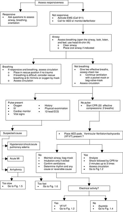
FIGURE 1.1. Universal Algorithm for Adult Emergency Cardiac Care AED, automatic external defibrillator; CPR, cardiopulmonary resuscitation; ECG, electro-cardiogram; EMS Emergency Management Service; IV, intravenous; MI, myocardial infarction

(

The American Heart Association in collaboration with the International Liaison Committee on Resuscitation: Guidelines 2000 for Cardiopulmonary Resuscitation and Emergency Cardiovascular Care Circulation 2000; 102: 8 Suppl I with permission; also adapted from Circulation 2005;Suppl III:1 130.

)

P.10


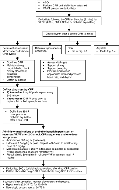
FIGURE 1.2. Algorithm for Ventricular Fibrillation (VF) and Pulseless Ventricular Tachycardia (VT) ABC, airway breathing circulation; CPR, cardiopulmonary resuscitation; IV, intravenous; PEA, pulseless electrical activity

Give drugs typically at 3 5 minute intervals: Vasopressin 40 U IV single dose (wait 10 minutes before giving epinephrine)

(

The American Heart Association in collaboration with the International Liaison Committee on Resuscitation: Guidelines 2000 for Cardiopulmonary Resuscitation and Emergency Cardiovascular Care. Circulation 2000; 102: 8 Suppl I, with permission; also adapted from Circulation 2005;Suppl III:1 130.

)

P.11


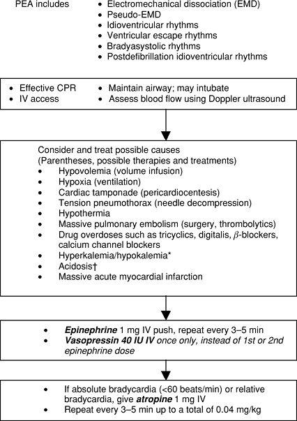
FIGURE 1.3. Algorithm for Pulseless Electrical Activity (PEA) CPR, cardiopulmonary resuscitation; EMD, electromechanical dissociation; IV, intravenous Class I: definitely helpful. Class IIa: acceptable, probably helpful. Class IIb: acceptable, possibly helpful. Class III: not indicated, may be harmful.

*Sodium bicarbonate 1 mEq/kg: is Class I: if patient has known pre-existing hyperkalemia.

Sodium bicarbonate 1 mEq/kg: Class IIa: if known pre-existing bicarbonate-responsive acidosis; if overdose with tricyclic antidepressants; to alkalinize the urine in drug overdoses. Class IIb: if intubated and long arrest interval; upon return to spontaneous circulation after long arrest interval. Class III: hypoxic lactic acidosis.

(

The American Heart Association in collaboration with the International Liaison Committee on Resuscitation: Guidelines 2000 for Cardiopulmonary Resuscitation and Emergency Cardiovascular Care. Circulation 2000; 102: 8 Suppl I with permission; also adapted from Circulation 2005;Suppl III:1 130.

)

P.12


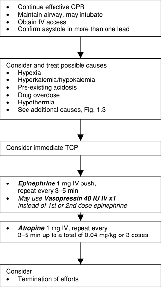
FIGURE 1.4. Asystole Treatment Algorithm CPR, cardiopulmonary resuscitation; IV, intravenous; TCP, transcutaneous pacing

(

The American Heart Association in collaboration with the International Liaison Committee on Resuscitation: Guidelines 2000 for Cardiopulmonary Resuscitation and Emergency Cardiovascular Care. Circulation 2000; 102: 8 Suppl I, with permission; also adapted from Circulation 2005;Suppl III:1 130.

)

P.13


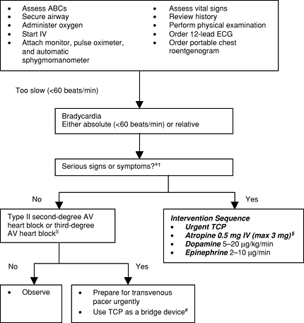
FIGURE 1.5. Bradycardia Algorithm (Patient not in Cardiac Arrest) ABC, airway breathing circulation; AV, atrioventricular; BP, blood pressure; ECG, electrocardiogram; HF, heart failure; IV, intravenous; MI, myocardial infarction; TCP, transcutaneous pacemaker

*Serious signs or symptoms must be related to the slow rate. Clinical manifestations include: symptoms (chest pain, shortness of breath, decreased level of consciousness) and signs (low BP, shock, pulmonary congestion, HF, acute MI).

Do not delay TCP while awaiting IV access or for atropine to take effect if patient is symptomatic.

Atropine should be given in repeat doses in 3 5 minutes up to total of 0.04 mg/kg.

Never treat third-degree heart block plus ventricular escape beats with lidocaine.

#Verify patient tolerance and mechanical capture. Use analgesia and sedation as needed.

(

The American Heart Association in Collaboration with the International Liaison Committee on Resuscitation: Guidelines 2000 for Cardiopulmonary Resuscitation and Emergency Cardiovascular Care. Circulation 2000; 102: 8 Suppl I, with permission; also adapted from Circulation 2005;Suppl III:1 130.

)

P.14


P.15


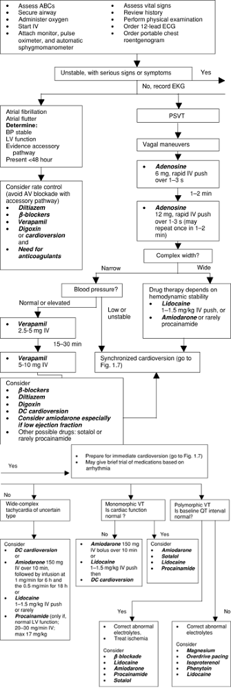
FIGURE 1.6. Tachycardia Algorithm ABC, airway breathing circulation; AV, atrioventricular; BP, blood pressure; DC, direct current; ECG, electrocardiogram; LV, left ventricular; PSVT, paroxysmal supraventricular tachycardia; VT, ventricular tachycardia

(

The American Heart Association in collaboration with the International Liaison Committee on Resuscitation: Guidelines 2000 for Cardiopulmonary Resuscitation and Emergency Cardiovascular Care. Circulation 2000; 102: 8 Suppl I, with permission; also adapted from Circulation 2005;Suppl III:1 130.

)

P.16


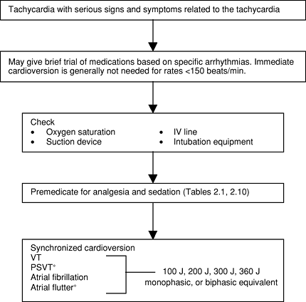
FIGURE 1.7. Electrical Cardioversion Algorithm (Patient not in Cardiac Arrest) IV, intravenous; VT, ventricular tachycardia

*PSVT (paroxysmal supraventricular tachycardia) and atrial flutter often respond to lower energy levels (start with 50 J)

(

The American Heart Association in collaboration with the International Liaison Committee on Resuscitation: Guidelines 2000 for Cardiopulmonary Resuscitation and Emergency Cardiovascular Care. Circulation 2000; 102: 8 Suppl I, with permission; also adapted from Circulation 2005;Suppl III:1 130.

)


Handbook of Critical Care Drug Therapy
ISBN: 0781797632
EAN: 2147483647
Year: 2006
Pages: 19

flylib.com © 2008-2017.
If you may any questions please contact us: flylib@qtcs.net